Direct Analysis of Phosphoproteins Using Mass Spectrometry
Bruker Application Note
Introduction
Protein phosphorylation is an essential mechanism in eukaryotic cells to regulate vital cellular processes such as cell cycle or growth and differentiation. Altered regulation may result in critical diseases like cancer. Detailed characterization is therefore essential. Phosphoproteins are challenging samples for direct analysis using mass spectrometry (MS). The instrument must be able to characterize the molecules against a high background of abundant proteins in the sample.
The new amaZon speed ETD ion trap MS is ready for this task. The instrument delivers a wide dynamic range, high spectral duty cycle and offers electron-transfer dissociation (ETD) fragmentation along with collision-induced dissociation (CID). By combining ETD with CID, the instrument is state-of-the-art in the characterization of phosphylation sites in proteins.
Experimental
A recent experiment by Tsai et al.1 analysed human Raji B cell lysates. In brief, cells were digested and phosphopeptide purification was performed. Vacuum-dried peptide samples were then separated using an Ultimate 3000 nano-LC system (Dionex, Sunnyvale, CA, USA) coupled to an amaZon speed ETD ion trap equipped with CaptiveSpray ion source (both Bruker, Bremen, Germany). Scan speed was 8,100 u/s in MS and 52,000 in MS/MS. Acquisition was performed with combined CID and ETD fragmentation. All results were stored and analysed with ProteinScape (Bruker).
Results and Discussion
In three technical replicates, an average of 320 proteins were identified. 313 proteins were identified as phosphoproteins. ProteinScape substantially simplified data analysis while CID in combination with ETD efficiently enabled mapping of phosphorylation sites on the majority of peptides studied. In [Figure 1(a)], phosphorylation of a peptide is identified using CID. ETD analysis is automatically triggered from the CID result. ETD fragmentation cleaves the peptide backbone, leaving the phosphate group in place and thus allows evaluation of the localization of the phosphate group [Figure 1 (b) and (c)] – in this case serine 3.


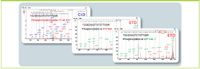
Figure 1: Characterization of phosphoproteins with CID and ETD: (a) CID identifies phosphorylated proteins, while ETD enables the location of the phosphate groups to amino acids (b and c). Here, serine 3 was identified as the phorsphorylation site.
Conclusions
ETD triggered by CID is by far the most efficient technology to characterize post-translational modifications. The experiments cited here clearly demonstrate the superior analytical power of the combination of state-of-the-art ion trap technology with optimized fragmentation techniques and leading ion source design. Powerful bioinformatics with ProteinScape enable comprehensive and reliable achievement of results.
Bruker Daltonik GmbH
Fahrenheitstr. 4, 28359 Bremen, Germany
tel: +49 421 2205 0 fax: +49 421 2205 104
E-mail: sales@bdal.de Website: www.bdal.com
References
1. Bruker Daltonics Technical Note # TN-42, Phosphoproteome mapping with the amaZon speed ETD. (www.bdal.com/tn-42).