Molecular Spectroscopy: Underutilized Detection for Gas Chromatography

Molecular spectroscopy techniques, most commonly ultraviolet-visible (UV-vis), are usually thought of as analyzing liquidphase samples, with techniques such as high-performance liquid chromatography (HPLC). Although not as common as in HPLC, molecular spectroscopy can be used with gas chromatography (GC) as well. In this installment, we examine molecular spectroscopy in combination with GC. The two most common techniques used with GC today are Fourier transform infrared spectrometry (FT-IR) and vacuum ultraviolet spectroscopy (VUV). Both provide structural and quantitative information and can be used in complementary fashion with the more common GC–mass spectrometry (GC–MS) and classical detectors. We will discuss the basics of GC–FT-IR and GC–VUV, when and when not to use them, and how they compare to and complement classical detectors.
n a recent installment, we discussed basic care and feeding common to all detectors for GC (1). We saw that there are myriad detectors, and that detection in capillary gas chromatography faces many challenges not seen in traditional spectroscopy. Analytes are in the vapor phase, generally at low (ppm–ppb) concentration, and are moving rapidly, often 50–100 cm/sec, as they are eluted from the column. A detector must have a rapid response, high sensitivity, and ruggedness. The vapor-phase analytes are often semivolatile; they must be maintained in the vapor phase as they exit the column, so the detector is heated to prevent condensation and deposition of the analyte. Chapters in books by Poole and McNair, Miller and Snow, and CHROMacademy, LCGC’s online training platform, provide overviews and details about detectors (2–4).
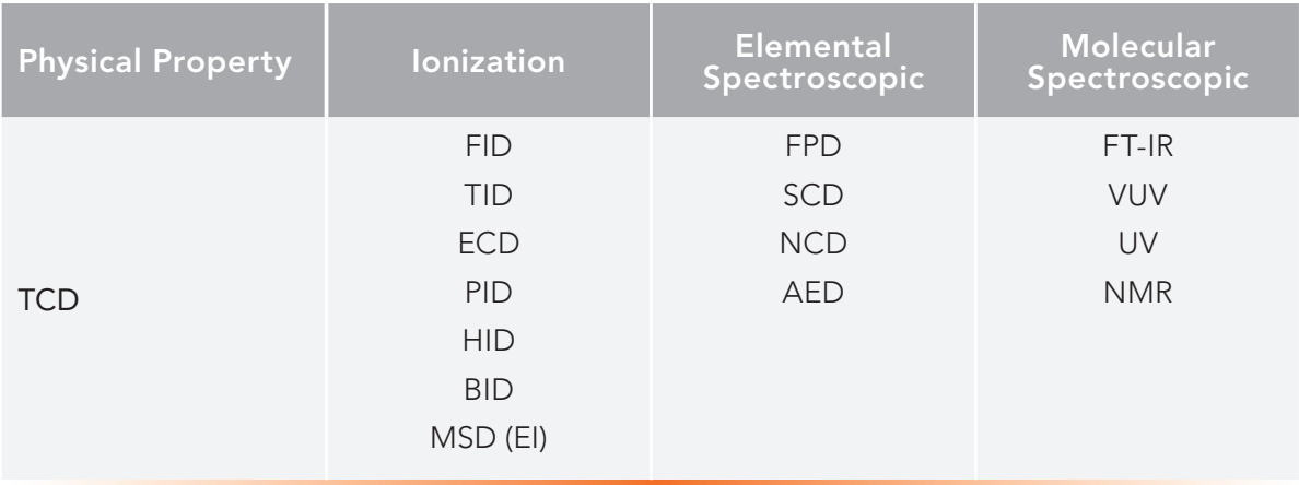
Table I provides a summary of many common detectors, grouped into four categories: physical property, ionization, elemental spectroscopic and molecular spectroscopic. The classical thermal conductivity detector (TCD), a physical property detector, is universal, detecting based on the difference in thermal conductivity between analytes and the background carrier gas. Ionization detectors, including electron ionization mass spectrometry (EI-MS) and flame ionization detection (FID), involve ionization of the analyte by various mechanisms and range from highly selective (electroncapture detection [ECD]) to nearly universal (FID). Spectroscopic detectors can be elemental, such as atomic emission (AED) or molecular. Elemental detectors often use high energy techniques to excite analytes. Like the classical spectroscopic detectors used in HPLC, molecular spectroscopic detectors for GC employ lower energy photons that do not ionize or decompose the analytes, and they behave in a very similar manner to their liquid-phase cousins, with a few exceptions. A detailed discussion and analysis of molecular spectroscopic detectors for GC is provided in a 2018 review by Zavahir, Nolvachai, and Marriott (5).


Table 1: Classification of some detectors by detection mechanism.
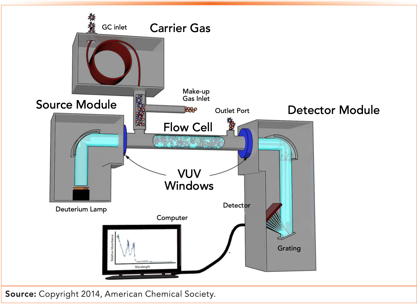
As in HPLC and classical spectroscopy, spectroscopic detectors obey the wellknown Beer-Lambert law, a form of which is shown in equation 1:


A is the absorbance of the analyte; a is the molar absorptivity of the analyte, a physical property dependent on the molecular structure, the wavelength of the incident radiation and the sample matrix; b is the pathlength of the cell or flow cell containing the analyte; and c is the concentration of the analyte. This presents challenges and opportunities particular to gas chromatography that are not seen in liquid-phase analytical techniques. First, the concentration of analyte in GC is necessarily low, as analytes are in the vapor phase and diluted with non-absorbing carrier gas. Low analyte concentration requires a longer pathlength, which provides the main challenge in using optical spectroscopic techniques with GC. The longer pathlength then results in extracolumn band broadening and some resulting loss of resolution and sensitivity.
The vapor-phase analysis required by GC also provides opportunities not easily seen in liquid- or solid-phase analysis. Detection in the vapor phase ensures clean spectra, with no interference from sample matrix or solvent, assuming that the chromatographic separation is complete. It also provides spectral detail and spectral range not generally available when a solvent is present, as we will especially see with vacuum ultraviolet (VUV) detection.
Spectroscopic detectors can provide a combination of qualitative and quantitative information. Infrared (IR) spectroscopy is a classical method for qualitative analysis; we all remember interpreting infrared spectra in our undergraduate training. IR generates signals through interaction of low-energy electromagnetic radiation that excite rotation and vibration of the chemical bonds in molecules. Ultraviolet (UV) spectroscopy has been implemented relatively recently with GC as VUV, using higher energy wavelengths, which excite high-energy electronic transitions. Traditional UV/visible (UV-vis) spectroscopy has been used with GC, but not widely (6). Interestingly, the two most common spectroscopic techniques used with GC straddle the traditional UV and visible range, with IR at lower energy and VUV at higher energy.
GC–VUV
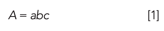
VUV operates in the same manner as traditional UV-vis, except at higher energy wavelengths, typically 120–240 nm, while classical UV-vis detectors used in HPLC typically operate from 200–800 nm. VUV gets its name from a classical need for a vacuum to reduce interferences from air, water, and background; however, today’s benchtop and instrument top detectors use a makeup gas to purge air and water, avoiding the need for a vacuum. A GC– VUV instrument operates like a classical UV-vis spectrometer, coupled to the GC. A diagram is shown in Figure 1 (7).

 detector coupled to a gas chromatograph. Reprinted with permission from reference (7).")
Figure 1: Diagram of a vacuum ultraviolet (VUV) detector coupled to a gas chromatograph. Reprinted with permission from reference (7).
In Figure 1, we see that effluent from the column exit is mixed with makeup gas and flows through a flow cell that serves the same role as the cuvette in a classical UV spectrophotometer. The other components are very similar to the classical instruments and detectors for HPLC. Incident radiation is provided by a modified deuterium lamp, which provides a broad spectrum of electromagnetic radiation to wavelengths as short as 120 nm. After traversing the flow cell, the light is focused onto a diffraction grating, which separates it by wavelength, then to a charge-coupled device (CCD) detector. This configuration allows rapid generation of high-quality UV spectra, enabling VUV as a rapid and highly selective detector.
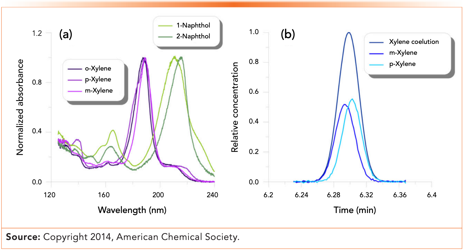
Figure 2 shows GC–VUV chromatograms of xylene and naphthol isomers. Note the spectral differences between the isomers, which make them easy to identify using a spectral library search. The left side shows the different spectra for the three xylene isomers and for two naphthol isomers. The right side shows a chromatogram of coeluted xylene isomers. Their distinct spectra make them easy to tell apart. In addition to providing qualitative spectral information, VUV also has promising figures of merit with limits of detection in picograms and 3–4 orders of magnitude linear range. It is also fast, with a response time of about 10 ms, making VUV amenable to fast and multidimensional GC.

 VUV spectra of xylene and naphthol isomers. (b) Chromatogram showing separation of the signals for m- and p-xylene. Reprinted with permission from reference (7).")
Figure 2: (a) VUV spectra of xylene and naphthol isomers. (b) Chromatogram showing separation of the signals for m- and p-xylene. Reprinted with permission from reference (7).
GC–FT-IR
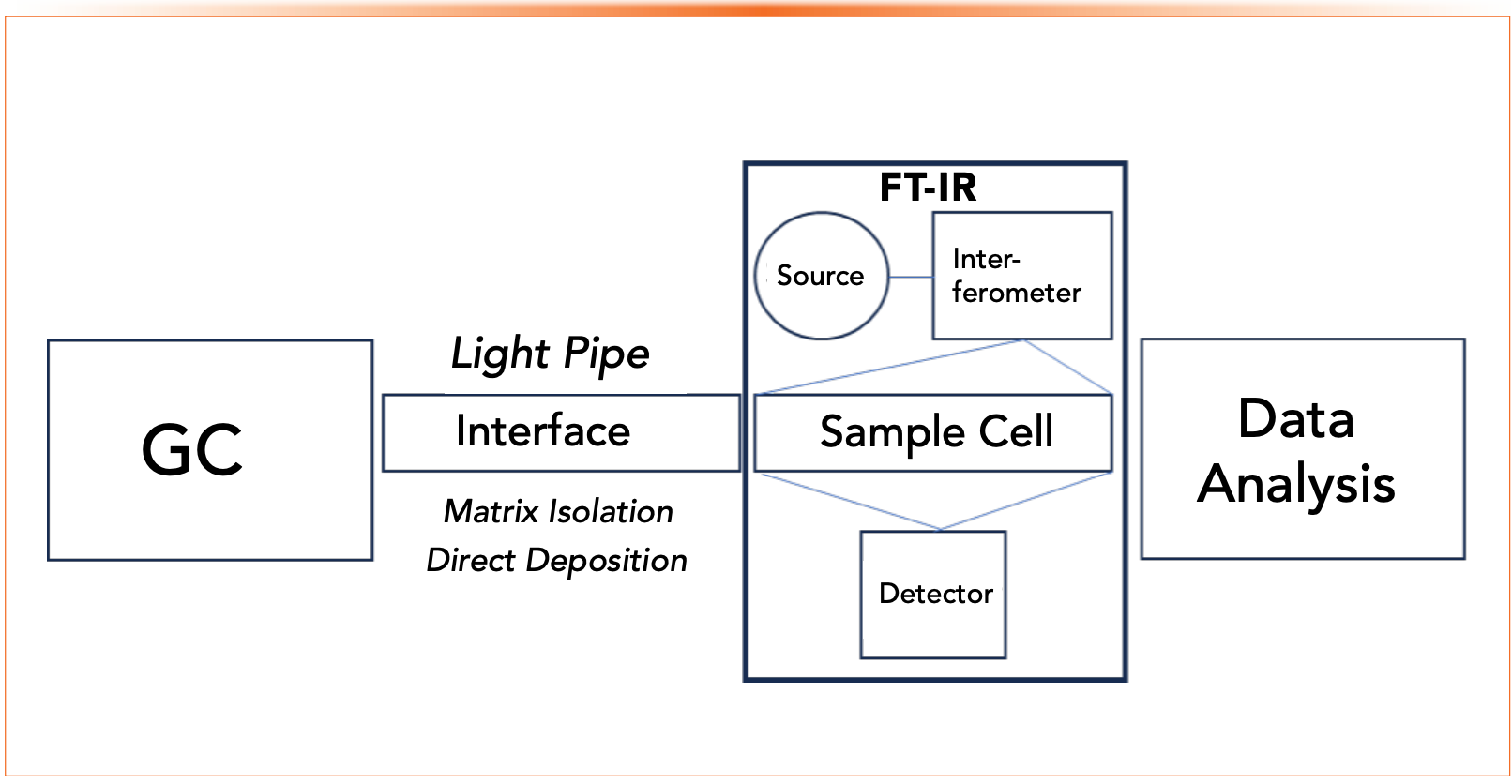
Nearly all chemists have used infrared spectroscopy, starting as undergraduates and finding it to be a powerful technique, especially in combination with other instrumental and chemical tests for structure determination. There is a long history of 50 or more years of work with GC coupled to FT-IR, yet due mostly to sensitivity concerns, GC–FT-IR remains a niche technique. Figure 3 shows a block diagram of a typical GC–FT-IR instrument. Like the VUV example, it is essentially a traditional spectrometer coupled to the outlet of the gas chromatographic column, with the same caveats discussed above for all detectors applied. The most common interface between GC and FT-IR is a light pipe, which is essentially a heated flow cell coupled to the end of the capillary column. Other interfaces include matrix isolation, in which the column effluent is cryogenically trapped under vacuum onto a gold-plated collection disc, and the related direct deposition, which places an IR microscope close to the column outlet. Matrix isolation and direct deposition provide greater sensitivity, while light pipe is much simpler.


Figure 3: Block diagram of a GC–FT-IR instrument showing basic components.
Complementary to mass spectrometry, FT-IR provides both universal and selective detection for organic analytes. Nearly all organic compounds absorb in the typical scanning range of IR spectrometers used with GC (4000–600 cm-1) providing universal detection, while scanning a spectrum provides unequivocal structural information. The combination of unique spectra, especially for structural isomers, and structural library searching, along with retention time data, allows highly selective qualitative analysis with GC–FT-IR.
Conclusions
While not as commonly used as ionization, elemental spectroscopic, or physical property detectors, molecular spectroscopic detectors, most commonly FT-IR and VUV, provide useful and complementary capabilities. They are non-destructive, so they can be used in tandem with other detectors. Both FT-IR and VUV are stars for qualitative analysis, especially for difficult problems in isomer analysis not easily addressed by MS. The longstanding problems with using FT-IR with GC, mostly having less sensitivity than MS, still exist and likely relegate FT-IR to a niche detector; however, there is sufficient sensitivity with relatively simple light pipe interfacing for many applications and FT-IR does not require some of the overhead of MS, such as high vacuum. VUV is a relatively new detector that offers sensitivity between FT-IR and MS, combined with a relatively simple instrument to operate and maintain, and excellent selectivity, especially for structural isomers not easily separated by MS. For problems involving structure determination with gas chromatography, mass spectrometry is not the only option; molecular spectroscopy offers benefits that make it worth considering.
References
(1) Snow, N. H. Basic Care and Feeding of Your Detector. LCGC N. Am. 2023, 41 (7), 256–258. DOI: 10.56530/lcgc.na.zd5789d1
(2) O’Brien, A. M.; Schug, K. A. Molecular Spectroscopic Detectors for Gas Chromatography. In Gas Chromatography, 2nd ed.; Poole, C. F., Ed.; Elsevier, 2021; pp 371–397. DOI: 10.1016/B978-0-12-820675-1.00021-6
(3) McNair, H. M.; Miller, J. M.; Snow, N. H. GC–MS and Spectrometric Detectors. In Basic Gas Chromatography, 3rd ed.; John Wiley & Sons, 2019; pp 157–175. DOI: 10.1002/9781119450795.ch10
(4) CHROMacademy. https://www.chromacademy.com/ (accessed 2023-10-18).
(5) Zavahir, J. S.; Nolvachai, Y.; Marriott, P. J. Molecular Spectroscopy – Information Rich Detection for Gas Chromatography. Trends Analyt. Chem. 2018, 99, 47–65. DOI: 10.1016/j.trac.2017.11.014
(6) Gras, R.; Luong, J.; Shellie, R. A. Direct Measurement of Trace Elemental Mercury in Hydrocarbon Matrices by Gas Chromatography with Ultraviolet Photometric Detection. Anal. Chem. 2015, 87 (22), 11429–11432. DOI: 10.1021/acs.analchem.5b02895
(7) Schug, K. A.; Sawicki, I.; Carlton, D. D. Jr.; et al. Vacuum Ultraviolet Detector for Gas Chromatography. Anal. Chem. 2014, 86 (16), 8329–8335. DOI: 10.1021/ac5018343
About the Author


Nicholas H. Snow is the Founding Endowed Professor in the Department of Chemistry and Biochemistry at Seton Hall University, and an Adjunct Professor of Medical Science. During his 30 years as a chromatographer, he has published more than 70 refereed articles and book chapters and has given more than 200 presentations and short courses. He is interested in the fundamentals and applications of separation science, especially gas chromatography, sampling, and sample preparation for chemical analysis. His research group is very active, with ongoing projects using GC, GC–MS, two-dimensional GC, and extraction methods including headspace, liquid–liquid extraction, and solid-phase microextraction. Direct correspondence to: LCGCedit@mmhgroup.com


TD-GC–MS and IDMS Sample Prep for CRM to Quantify Decabromodiphenyl Ether in Polystyrene Matrix
April 26th 2024At issue in this study was the certified value of decabromodiphenyl ether (BDE 209) in a polystyrene matrix CRM relative to its regulated value in the EU Restriction of Hazardous Substances Directive.

LC–MS/MS-Based System Used to Profile Ceramide Reactions to Diseases
April 26th 2024Scientists from the University of Córdoba in Córdoba, Spain recently used liquid chromatography–tandem mass spectrometry (LC–MS/MS) to comprehensively profile human ceramides to determine their reactions to diseases.

High-Throughput 4D TIMS Method Accelerates Lipidomics Analysis
April 25th 2024Ultrahigh-pressure liquid chromatography coupled to high-resolution mass spectrometry (UHPLC-HRMS) had been previously proposed for untargeted lipidomics analysis, but this updated approach was reported by the authors to reduce run time to 4 min.


