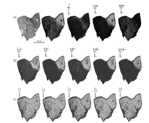
Over the last decade, matrix-assisted laser desorption–ionization (MALDI) imaging has become an indispensable tool for a broad range of applications, from studying plant metabolomics to discovering biomarkers of disease to developing new therapies. As such, MALDI imaging is revolutionizing preclinical drug discovery pipelines by providing direct distribution monitoring of therapeutic compounds and their metabolites along with untargeted pharmacodynamic information. A key application of MALDI imaging is tissue analysis for oncology, and recent developments in MALDI technology promise greater benefits to cancer research. The combination of MALDI with laser-induced post-ionization (PI) enhances the detection and imaging of pharmaceutical compounds and other classes of compounds, allowing for significant advances in the use of MALDI imaging for studying drug metabolism and pharmacodynamics in tumor tissues. This article describes the value of MALDI Imaging for oncology applications and examines the potential for laser-induced PI, including the ability to achieve up to three orders of magnitude higher sensitivity and to image metabolite classes previously undetectable with traditional MALDI.


