Skin Cells Tested Using MALDI-MSI Techniques
A group of scientists from the Luxembourg Institute of Science and Technology (LIST) in Belvaux, Luxembourg recently tested matrix-assisted laser desorption/ionization (MALDI) mass spectrometry imaging (MSI) to analyze human skin cells. Their work was published in the Journal of the American Society for Mass Spectrometry (1).
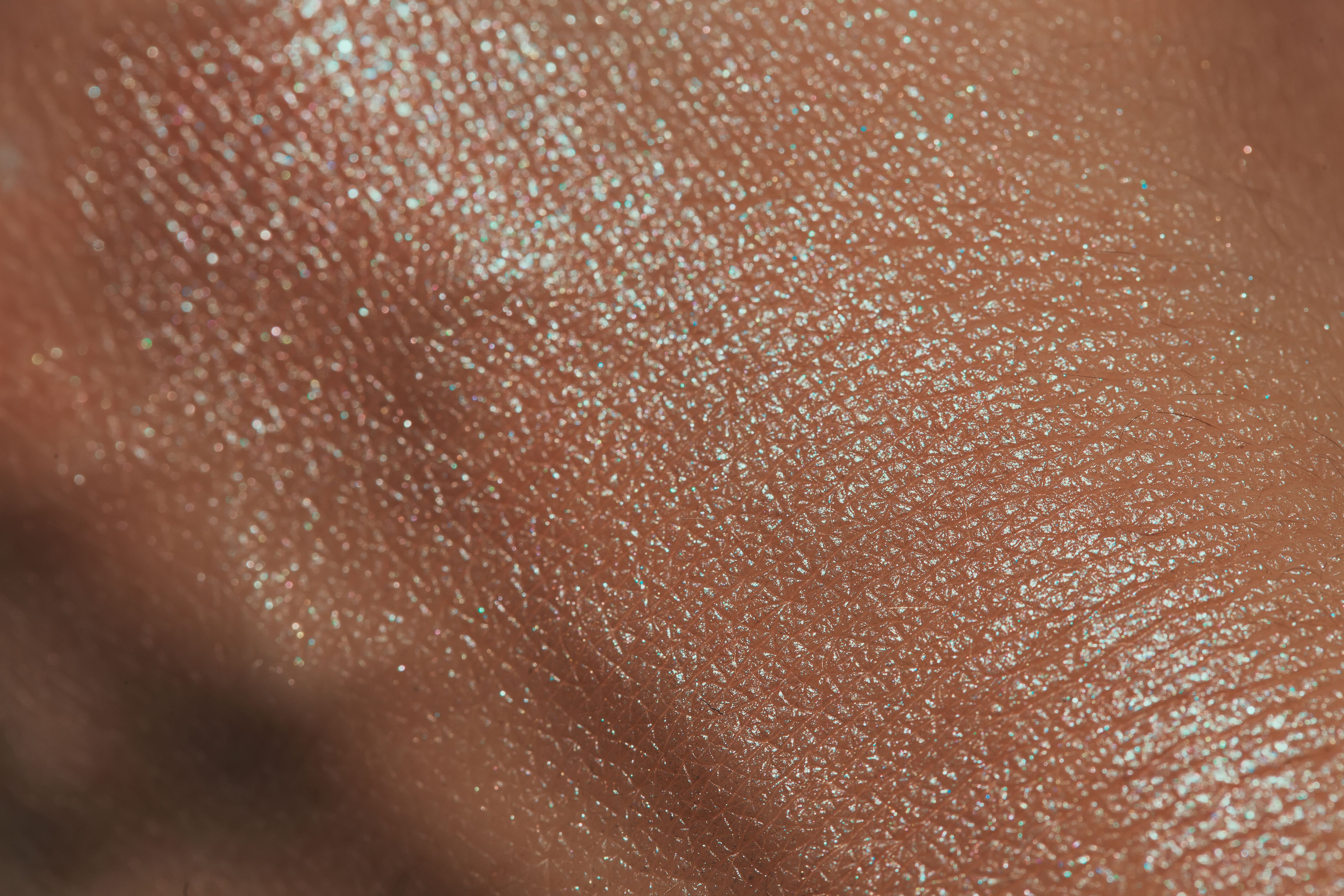
texture of human skin with liquid highlighter swatch | Image Credit: © viki2103stock - stock.adobe.com

One technique used to analyze skin and either search for disease or conduct biological research is MALDI coupled with MSI. MALDI is a soft ionization technique that uses an energy-absorbing matrix to aid in analyte detection; when combined with MSI, which can enable investigations into the spatial distribution of molecular species in different samples (2,3), it can be effective in different analysis scenarios. When it comes to measuring skin penetration and local skin toxicity, usual materials include in vivo animal models and ex vivo animal skin; however, due to high cost, structural differences between human and animal skin, and ethical issues, scientists are searching for better ways to test. Reconstructed human epidermis (RHE) models can be used to mimic human skin and analyze chemicals’ adverse effects on skin. However, there are few studies investigating the effectiveness of MALDI–MSI on RHE.
To determine the potential of this technique, multiple factors, such as different substrates, sample thickness, and tissue treatments, such as washing and recrystallization for fixed matrix parameters, were tested on the MALDI-MSI technique. This technique was then applied to lipids analysis of the SkinEthic RHE model. Images were generated using an atmospheric pressure MALDI source coupled to a high-resolution mass spectrometer with a 5µm pixel size. From there, masses detected within a region of interest were analyzed and annotated using the LipostarMSI platform.
Three factors were identified as leading to images with significant signal intensities in addition to numerous m/z values: coated metallic substrates, such as APTES-coated stainless-steel plates; tissue sections of 6 μm thickness; and aqueous washing before HCCA matrix spraying (without recrystallization). This resulting data, according to the scientists, should improve sample preparation workflows for evaluating changes in skin composition post dermato-cosmetic application.
References
(1) Feucherolles, M.; Le, W.; Bour, J.; Jacques, C.; Duplan, H.; Frache, G. A Comprehensive Comparison of Tissue Processing Methods for High-Quality MALDI Imaging of Lipids in Reconstructed Human Epidermis. J. Am. Soc. Mass Spectrom. 2023, 34 (11), 2469–2480. DOI: https://doi.org/10.1021/jasms.3c00185
(2) MALDI Basics. Shimadzu Scientific Instruments 2023. www.ssi.shimadzu.com/service-support/faq/maldi/index.html (accessed 2023-11-10)
(3) Buchberger, A.R.; DeLaney, K.; Johnson, J.; Li, L. Mass Spectrometry Imaging: A Review of Emerging Advancements and Future Insights. Anal. Chem. 2018, 90 (1), 240–265. DOI: 10.1021/acs.analchem.7b04733
TD-GC–MS and IDMS Sample Prep for CRM to Quantify Decabromodiphenyl Ether in Polystyrene Matrix
April 26th 2024At issue in this study was the certified value of decabromodiphenyl ether (BDE 209) in a polystyrene matrix CRM relative to its regulated value in the EU Restriction of Hazardous Substances Directive.
LC–MS/MS-Based System Used to Profile Ceramide Reactions to Diseases
April 26th 2024Scientists from the University of Córdoba in Córdoba, Spain recently used liquid chromatography–tandem mass spectrometry (LC–MS/MS) to comprehensively profile human ceramides to determine their reactions to diseases.
High-Throughput 4D TIMS Method Accelerates Lipidomics Analysis
April 25th 2024Ultrahigh-pressure liquid chromatography coupled to high-resolution mass spectrometry (UHPLC-HRMS) had been previously proposed for untargeted lipidomics analysis, but this updated approach was reported by the authors to reduce run time to 4 min.