

News|Articles|September 30, 2026 (Updated: September 30, 2026)
Chromatography Boosts EV Isolation from Plasma
Author(s)John Chasse
Size exclusion chromatography (SEC) plus precipitation improves extracellular vesicle (EV) purity from blood plasma.
Advertisement
Extracellular vesicles (EVs) are tiny, membrane-covered particles that cells release into body fluids like blood, saliva, and urine, both in normal conditions and during disease. They're seen as a promising tool for detecting disease because they carry cargo (DNA, RNA, proteins, and fats) that reflects what is happening inside the cells that made them.
The challenge is that pulling EVs out of blood plasma is technically difficult due to plasma being packed with lipoproteins and other proteins that are similar in size and density to EVs, making them hard to separate out. Several methods are commonly used to isolate EVs, including ultracentrifugation (UC) which spins samples at very high speeds, size exclusion chromatography (SEC) which filters by size, and precipitation (Precip), which chemically causes EVs to clump and settle out. None of these methods, however, can completely get rid of the non-EV material that tags along.
To try to solve this problem, researchers tested two approaches that combined different isolation methods together (UC followed by Precip and SEC followed by Precip) to get more EVs out of blood plasma while also making them cleaner and less contaminated. To thoroughly check the quality of the EVs they isolated, the researchers looked at several things: how much protein and fat they contained, the range of particle sizes present, and how pure the samples were. A paper based on their efforts was published in the Journal of Biotechnology.1
Why is Blood Plasma a Valuable but Challenging Source for Isolating EVs?
Among the body fluids that contain EVs, blood plasma is one of the richest sources. Drawing blood is a routine procedure that rarely causes patients much discomfort, making it a practical way to collect samples for further analysis. Blood plasma is especially useful because it also carries other biochemical information that's already commonly used in clinical care. Because of this, EVs found in plasma can offer valuable clues about how various diseases are likely to progress, helping doctors choose the right treatments and preventive steps. Overall, collecting EV samples this way is simple, quick, and minimally invasive. That said, plasma is a complicated fluid, which makes isolating EVs from it difficult. It contains many dissolved proteins, like albumin and globulins, along with different types of fat-carrying lipoproteins—mainly chylomicrons, high-density lipoprotein (HDL), low-density lipoprotein (LDL), intermediate-density lipoprotein (IDL), and very low-density lipoprotein (VLDL).2 In addition, these lipoproteins share similar features with EVs—like size and density—which makes it even harder to tell the two apart and separate them.2,3 “Therefore,” write the authors of the paper,1 “obtaining high purity EV samples from plasma without co-isolating non-EV contaminants is difficult.”
Which Combined Isolation Method Works Best for Obtaining High-Quality EVs from Blood Plasma?
The results of this study showed that combining SEC with a precipitation chemical—especially one called ExoQuick-TC—kept the EVs intact and the samples clean. On the other hand, combining UC with precipitation didn't work as well: it captured fewer EVs and needed much larger amounts of blood plasma to work, which makes it less practical for use in real clinical settings.1
“Overall,” write the authors of the paper,1 “our study suggests that SEC followed by Precip is a suitable strategy for obtaining EV-enriched plasma samples compatible with downstream characterization and biomarker studies.”
Read More on Similar Topics
References
- Serrano-Pertierra, E.; Álvarez, E. S.; Blanco-López, M. C. et al. Enhancing Extracellular Vesicles Purity from Plasma: A Comparative Study of Isolation Strategies. J Biotechnol. 2026, S0168-1656 (26), 00252-X DOI:
10.1016/j.jbiotec.2026.09.006 - Sódar, B. W.; Kittel, Á.; Pálóczi, K. et al. Low-density Lipoprotein Mimics Blood Plasma-derived Exosomes and Microvesicles During Isolation and Detection. Sci. Rep.2016, 6 (1), 24316. DOI:
10.1038/srep24316 - Menard, J. A.; Cerezo-Magaña, M.; Belting. M. Functional Role of Extracellular Vesicles and Lipoproteins in the Tumour Microenvironment. Philos. Trans. R. Soc. B Biol. Sci. 2018, 373 (1737), 20160480. DOI:
10.1098/rstb.2016.0480
Advertisement
Related to this article
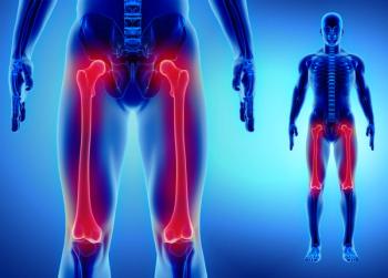
Data-independent acquisition (DIA) liquid chromatography-tandem mass spectrometry (LC–MS/MS) reveals shared and site-specific proteins across the thighbone.
Laura Righetti explains why quantification, not instrumentation, still keeps mass spectrometry imaging (MSI) out of routine food safety testing.

Large language models help chromatographers build LC pressure troubleshooting apps in hours.

Magnetic molecularly imprinted polymer extraction with liquid chromatography-high resolution mass spectrometry (LC–HRMS) finds patulin in store-bought juice.

Richard Bade and Malcolm McLeod explain to LCGC International how liquid chromatography-tandem mass spectrometry (LC-MS/MS) detects drug trends in wastewater.

In this fifth part of " Conversations with Michael Dong," a series on fundamentals of HPLC in pharmaceutical analysis, LCGC International’s "Perspectives in HPLC" column editor discusses how to overcome challenges with reversed-phase chromatography in drug analysis.
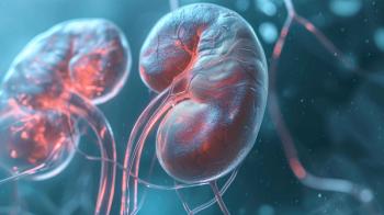
Liquid chromatography–tandem mass spectrometry (LC–MS/MS) and microbiome data reveal kidney markers of diabetic disease.
Advertisement
Advertisement
Trending on LCGC International
1
LC–MS/MS Tracks Drugs in Australian Wastewater
2
LC–HRMS and Magnetic Traps Detect Patulin in Apple Juice
3
A New Era in LC Troubleshooting Tools
4
Mass Spectrometry Imaging in Food: The Power and the Limits
5