- The Column-11-06-2019
- Volume 15
- Issue 11
Diagnosing Cancer Using Cerumen and HSGC–MS
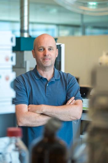
The Column spoke to Nelson Roberto Antoniosi Filho, a professor at the Chemistry Institute of the Federal University of Goiás (UFG), in Goiânia, Brazil, about his development of a gas chromatography–mass spectrometry (GC–MS) method for cancer diagnosis using cerumen.
The Column spoke to Nelson Roberto Antoniosi Filho, a professor at the Chemistry Institute of the Federal University of Goiás (UFG), in Goiânia, Brazil, about his development of a gas chromatography–mass spectrometry (GC–MS) method for cancer diagnosis using cerumen.
Q. In a recent study, your group developed a new method using cerumen as a biomarker for cancer diagnosis (1). What led your group to begin this study?
A: The idea of analyzing the chemical composition of earwax emerged in the 1990s when I was a Ph.D. student performing chemical analysis of oils and fats by gas chromatography (GC) and computational methods. The intention was to know the composition of other matrices that supposedly had lipids in their chemical compositions.
The application for clinical diagnosis of diseases appeared in 2014 when I was approached by Egyptian researcher, Engy Shokry, to pursue postdoctoral studies under his supervision. Engy had experience in the diagnosis of diseases by blood analysis and I thought it would be interesting to also evaluate the possibility of diagnosing diseases by analyzing earwax, because it is a product of metabolic secretion.
The first work developed was the diagnosis of Diabetes mellitus types I and II (2), which led to me proposing the study of a cancer diagnosis using the same methodology developed for the diagnosis of diabetes. During this period, several other works in the pharmaceutical (3), medicinal (4), and veterinary areas (5,6) were developed, demonstrating the potential of earwax to be a fingerprint of what happens in the cells of living beings (1).
Q. The early identification and diagnosis of cancer is paramount to a successful outcome. The noninvasive nature of the matrix collection can only serve to help this. Why did you specifically choose to analyze cerumen?
A: From a medical point of view, cerumen is formed by two glands called apocrine, which, from a chemical point of view, seem to secrete polar and nonpolar substances. This means that cerumen, despite being secreted in small quantities, is a concentrated and diverse mix of chemical substances that, as we observed, reflect what happens in our metabolism. In addition, we noted that regardless of the type of cancer, there are chemical substances that appear in all types of cancers and can identify the occurrence of this disease; in the cerumen these substances appear even in the early stages of the disease, allowing early diagnosis, which will decrease the mortality and the emotional and financial losses associated with the disease. Thus, cerumen seems to be the best fingerprint of what happens in our body, which perhaps will make this biomatrix the best option for the diagnosis of many other diseases, not just cancer, diabetes, and other diseases that we have already verified can be identified (results not yet published) with this technique, which we call cerumenogram.
Q. Why was headspace gas chromatography–mass spectrometry (HSGC–MS) your technique of choice? What does this technique offer over others?
A: The HSGC–MS technique was chosen because it allows the analysis of volatile compounds without the need for any sample preparation steps. In addition, it allows the emission of volatile organic compounds (VOCs) from earwax to be increased, increasing the amount and diversity of biochemical information provided by this biological secretion, which allows excellent discriminations to be observed between healthy and cancer individuals. In addition to its practicality and richness of information, the HSGC–MS technique is one of the most financially accessible extraction, chromatographic separation, and spectrometric identification techniques, and is therefore easily found in many parts of the world, which will help to disseminate the use of the cerumenogram.
Q. Did the waxy nature of cerumen present any difficulties for analysis?
A: Not at all. The headspace extraction simplifies the analysis of volatile and semivolatile compounds, with no prior sample preparation, and we did not observe any carryover effects in the syringe or on the chromatography column during the clean steps or blanks analyses.
Q. Can you tell us more about the multivariate analysis stage?
A: The cerumenogram leads to a total ion chromatogram (TIC) with too many peaks. Hence, the first step is the application of GC–MS peaks quality check-up: baseline correction, noise reduction, retention time alignment, and data-normalization. In total, 158 peaks were marked as cerumen metabolites. Previous studies have demonstrated that the concentrations of volatile compounds in cerumen are altered by some factors associated with sex/gender and ethnicity/race. For this reason, we transform our data matrix into a binary output (absence/presence) and run statistical analysis to observe if the factors associated with sex/gender and ethnicity/race of the individual is an influence in the discrimination of our data set. Fortunately, when the binary approach is applied, there is no discrimination due to sex/gender and ethnicity/race of the individuals (as can be observed in the supplementary information of our paper). Moreover, to select the cancer biomarkers among the 158 volatile organic metabolites (VOMs) identified, we have used tools of the evolutionary algorithms (EAs), the genetic algorithm for a partial least squares regression (GA-PLS). The GA-PLS selected 27 metabolites as potential cancer biomarkers present in cerumen. Aiming to confirm if the compounds selected by GA-PLS were capable to distinguish between cancer and non-cancer individuals, an unsupervised machine learning method-hierarchical cluster analysis (HCA)-was carried out with the original outputs of the 27 VOMs selected, providing a 100% discrimination between cancer and healthy individuals.
Q. What is novel about the method you developed?
A: The application of cerumen as a useful biofluid for cancer diagnoses for the first time and the observation that there are some VOCs that are commonly produced by all types of cancer. As well, the ease of the diagnoses, since the sample collection is easy, noninvasive, and painless. In addition, HSGC–MS appears as a new, practical, quick, robust, effective, and simple way to provide an accurate diagnosis of an important disease normally diagnosed by painful and expensive methods.
Q. What were your main findings?
A: One of the main findings was the highest accuracy possible in the sample discrimination of cancer and healthy individuals. Our analysis, cerumenogram, provides 100% effectiveness in cancer identification based on the data obtained. We have also analyzed samples from individuals in the first stage of cancer, and our method was able to identify cancer in early stages, which is extremely important to increase the chances of a successful outcome.
Q. What are the next steps to be taken with your results?
A: Our next step aims to increase the number of test individuals for the development of new statistical models. In this way, we look for reach in not only cancer detection but also the identification of cancer type and its stages. We also intend to perform studies about the biochemical pathways of the cancer biomarkers in cerumen, with the purpose of better understanding how they get into this biomatrix and if its blockade may be helpful in cancer regression. In addition, we are also evaluating the applicability of cerumenogram in the diagnosis of other metabolic disorders, such as autism, depression, Alzheimer’s disease, xeroderma pigmentosum, and Parkinson’s disease.
References
- J.M.G. Barbosa, N.Z. Pereira, L.C. David, C.G. de Oliveira, M.F.G. Soares, M.A.G. Avelino, A.E. de Oliveira, E. Shokry, and N.R. Antoniosi Filho, Scientific Reports9, 11722 (2019). https://doi.org/10.1038/s41598-019-48121-4
- E. Shokry, A.E. de Oliveira, M.A.G. Avelino, M.M. de Deus, and N.R. Antoniosi Filho, Journal of Proteomics159, 92–101 (2017).
- E. Shokry, J.G. Marques, P.C. Regazzo, N.Z. Pereira, and N.R. Antoniosi Filho, Forensic Toxicology35(2), 348–358 (2017).
- E. Shokry, A.E. de Oliveira, M.A.G. Avelino, M.M. de Deus, N.Z. Pereira, and N.R. Antoniosi Filho, Forensic Toxicology35(2), 389–398 (2017).
- E. Shokry, F.C. Dos Santos, P.H.J da Cunha, M.C.S. Fioravanti, A.D.F. Noronha Filho, N.Z. Pereira, and N.R. Antoniosi Filho, Toxicon. 137, 54–57 (2017).
- E. Shokry, J. Pereira, J.G. Marques Junior, P.H.J da Cunha, A.D.F. Noronha Filho, J.A. da Silves, M.C.S. Fioravanti, A.E. de Oliveria, and N.R. Antoniosi Filho, PLoS ONE12(8), e0183538 (2017).
Nelson Roberto Antoniosi Filho is a professor at the Chemistry Institute of the Federal University of Goiás (UFG) where he coordinates the Laboratory of Extraction and Separation Methods (LAMES) in the development of analytical methodologies for the analysis of fuels, foods, natural products, drugs, pollutants, and biomatrices.
E-mail:
Articles in this issue
about 6 years ago
Agilent Opens New UK Spec Facilityabout 6 years ago
SepSolve Analytical Unveils New Canadian Facilityabout 6 years ago
Challenging Allergen Uncertainty Using LC–MS/MSabout 6 years ago
Sartorius Announces Acquisition of Danaher Life Science Portfolioabout 6 years ago
Analysis of White Oils Using AgLC×GCabout 6 years ago
Introduction to Glycan Analysisabout 6 years ago
Vol 15 No 11 The Column November 2019 North American PDFNewsletter
Join the global community of analytical scientists who trust LCGC for insights on the latest techniques, trends, and expert solutions in chromatography.


