
|Articles|October 7, 2022
- October 2022
- Volume 18
- Issue 10
- Pages: 16 – 21
Modern Size-Exclusion Chromatography Separations of Biosimilar Antibodies at Physiological pH and Ionic Strength
Author(s)Stephan Koza, Bill Warren
Advertisement
The state of protein-derived self-associated, aggregated and fragmented impurities in biotherapeutics are critical quality attributes (CQAs) and are widely monitored using non-denaturing size-exclusion chromatography (SEC). The mobile phases often used for accurate quantification of impurities on high sample-throughput SEC columns often deviate from the pH of the formulation buffer and can be of much higher ionic strength, which may result in the dissociation of non-covalently aggregated proteins during SEC analysis. Therefore, it may be beneficial to use phosphate buffered saline (PBS) at a physiologically relevant pH and ionic strength as an SEC eluent for the analysis of monoclonal antibody size variants (mAbs).
One of the major limitations in conducting reliably accurate size‑exclusion chromatography (SEC) of various monoclonal antibody (mAb) biotherapeutic aggregates, desired monomers, and fragments is undesired secondary interactions stemming from a lack of SEC particle and/or column hardware inertness. Consequently, scientists have had to invest significant time and resources to determine the effect that SEC eluent pH and ionic strength have on these protein size variant separations. In addition, scientists have desired yet struggled to find a “generic/platform SEC eluent formulation” for SEC columns for the ultrahigh‑performance liquid chromatography (UHPLC) or high performance liquid chromatography (HPLC) -based SEC analysis of various mAbs, including biosimilars and antibody–drug conjugates (ADCs).
Cross-linked dextran-agarose SEC particles (> 8.6 µm) in glass columns have been commonly used for the analysis of proteins at or near physiological conditions for over 30 years because they exhibit superbly low levels of unspecific ionic and hydrophobic interactions (1). However, the comparatively larger particle size and the compressibility of dextran-agarose SEC particles require the use of lower operating flow rates compared with the use of more rigid silica-based SEC particles. This results in longer analysis times to obtain desired mAb aggregate, monomer, and fragment resolution and quantification.
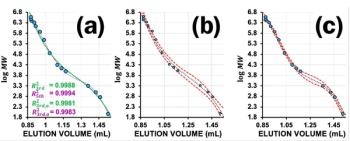
Chromatographers who perform SEC for mAb size variant analysis have frequently expressed a desire to use a single SEC eluent, for example, a platform SEC eluent/method, to obtain accurate and reproducible data for these determinations. Ideally, the ability to use commercially available, filtered, and sterile phosphate buffered saline (PBS) would significantly reduce the time to make as well as test these different SEC eluent formulations. Furthermore, the use of PBS as an SEC mobile phase provides a pH and tonicity consistent with serum, interstitial fluid, and lymph, which are primary fluids that an intravenously or subcutaneously administered parenteral therapeutic protein is exposed to, making it a relevant SEC eluent choice. However, as shown in Figure 1, significant method development resources evaluating the independent effects of SEC eluent pH and ionic strength are frequently required to obtain the desired results of biosimilar mAbs, ADCs, and other mAb derivatives, such as bi- or tri-specific antibodies.
SEC column manufacturers have actively worked to create new SEC particle and column hardware technologies that minimize undesired secondary ionic and hydrophobic interactions between proteins and many UHPLC and HPLC SEC column offerings (2,3). As shown in Figure 2, the synergistic combination of BEH SEC particles bonded with hydroxy-terminated polyethylene oxide (PEO), efficiently packed in “inert” stainless steel column hardware, delivers desired mAb aggregate, monomer, and fragment component resolution throughout a wide range of SEC eluent pH and ionic strengths. The purpose of the SEC column surface hardware modification is to greatly reduce ionic interactions that occur with the formation of meta‑complexes between the analyte proteins and metallic surfaces while maintaining the high efficiency and reproducible SEC particle packing capabilities obtained when using traditional stainless steel column hardware. In addition, the diol bonding of the BEH SEC particle has been supplanted by PEO to further minimize non-desired secondary hydrophobic interactions.
Method
To take advantage of the desired properties of “inert” UHPLC and HPLC column surface hardware, the separation of four commercially available biosimilars using PBS (1.15 g/L anhydrous Na2HPO4, 0.2 g/L KH2PO4, 0.8g/L NaCl, 0.2 g/L KCl, pH 7.4) SEC mobile phases at ionic strengths ranging from 150 mM (1× PBS), 120 mM (0.8× PBS), and 300 mM (2× PBS) were evaluated on 2.5 µm and 1.7 µm Waters MaxPeak Premier Protein 250 Å SEC columns. In addition, the commercially available cross-linked, dextran-agarose SEC column (Superdex 200 Increase 10/300GL, particle size >8.6 µm) (Cytiva) was also included due to the known “inert” properties of these SEC particles packed in glass columns. Examples of the mAbs that are currently available commercially in the United States as biosimilars are bevacizumab (25 mg/mL), infliximab (10 mg/mL), rituximab (10 mg/mL), and trastuzumab (21 mg/mL). All samples were analyzed neat following one or more freeze-thaw cycles that were performed to increase potential mAb aggregate formation. Note that as shown in Figure 1, a 7.8 × 300 mm, 2.5 µm mAb SEC 200 Å column (column 1) (Waters: BEH-DIOL SEC particles in stainless steel hardware) was also evaluated, and compared with separation performed on a 7.8 × 300 mm, 2.5 µm XBridge Premier Protein SEC 250 Å column (Waters: BEH-PEO SEC particles in “Inert” column hardware), to look at the influence of SEC eluent pH and ionic strength on the separation of the mAb trastuzumab.
For this work, the dextran-agarose SEC column was run at a flow rate of 0.25 mL/min to maximize resolution. Other scientists have reported that despite the low levels of protein-column interactions afforded using dextran-agarose SEC particles in glass columns, some protein size variants are not fully recovered due to unspecific ionic interactions (4).
Therefore, these same four biosimilar mAbs were evaluated on a dextran-agarose SEC column at PBS concentrations ranging from 0.8× to 2×, with collected data demonstrating reproducible recoveries (data not shown). As a result, and in the absence of additional corroborative data such as analytical ultracentrifugation (AUC), the high-molecular-weight species (HMWS 1 and HMWS 2) content observed using the Superdex 200 Increase 10/300GL 8.6 μm SEC column was used to evaluate the effectiveness and recoveries on a 7.8 × 300 mm, 2.5 μm XBridge Premier Protein 250 Å SEC column (Waters) and a 4.6 × 300 mm, 1.7 μm Acquity Premier Protein 250 Å SEC column (Waters). The flow rate used for the 2.5 μm BEH-PEO column was 0.5 mL/min as previously described, while the flow rate for the 1.7 μm BEH‑PEO column was 0.35 mL/min, which is twice the linear velocity used for the XBridge column and was possible due to the higher packed column efficiency of the 1.7 μm versus the 2.5 μm columns. As a result, the analysis times for the dextran‑agarose SEC column, 2.5 μm BEH‑PEO column, and 1.7 μm BEH‑PEO column were approximately 63 min, 16 min, and 12.5 min, respectively.
Results
Consistent chromatographic profiles for the biosimilar mAbs (Figure 3) and comparable quantitative results for HMWS1 and HMWS2 (Figure 4) were obtained on the dextran-agarose 8.6 μm in glass, as well as BEH-PEO 2.5 μm and BEH-PEO 1.7 μm particles contained in “inert” stainless steel hardware, with the predominant differences in component resolutions resulting from differing SEC column efficiencies.
These results indicate that the surface chemistry of the inert column hardware and BEH-PEO particles have reduced unspecific interactions with the four biosimilar mAbs in comparison with results obtained using the previous generation of BEH-diol-bonded SEC particles contained in unmodified stainless steel column hardware. More specifically, these results correlate with the literature‑derived isoelectric points (pI) of these mAbs. The reported measured pI values of trastuzumab (pI = 9.1) and rituximab (pI = 9.4) are significantly more basic than bevacizumab (pI = 8.3) and infliximab (pI = 7.6), which may account for the greater degree of observed non-desired cationic interaction at pH 7.6, with low abundance negative charges such as silanol-diol or BEH-diol SEC particles in packed SEC columns (5).
These results indicate that compared with many silica-diol or BEH-diol SEC particles contained in stainless steel hardware, BEH‑PEO SEC particles contained in “inert” stainless steel hardware provide a broader range of SEC eluent formulations capable of delivering effective protein size variant separations (biosimilar mAbs) and, as such, should be more amenable for use in platform SEC analytical methods.
By reducing the flow rate on the dextran‑agarose SEC column to 30% of the linear velocity used on the SEC column containing BEH-PEO 2.5 μm particles contained in “inert” stainless steel hardware, an effective separation of the HMWS variants was obtained, while the low-molecular-weight species (LMWS 1) fragments were partially resolved.
However, comparative run times on the dextran-agarose SEC column were increased approximately fourfold.
Conclusion
These results demonstrate that effective and robust high sample throughput SEC separations for four biosimilar mAbs were achieved using a 1× PBS physiological buffer (20 mM phosphate buffer with 150 mM NaCl, pH 7.4) as an SEC mobile phase on dextran-agarose 8.6 μm, BEH‑PEO 2.5 μm, and BEH-PEO 1.7 μm particles contained in glass or “inert” stainless steel column hardware, respectively. While it is not expected that PBS will be an effective mobile phase for the SEC analyses of all protein size variants, the use of PBS at varying concentrations offers the potential for simpler method development, transfer, and ease of use for mAb biosimilars and other proteins where reliable and accurate size‑based separations are needed.
References
- T. Arakawa, D. Ejima, T. Li, and J.S. Philo, Journal of Pharmaceutical Sciences 99(4), 1674–1692 (2010).
- T.H. Walter, B.A. Alden, J.L. Belanger, K. Berthelette, C. Boissel, M. DeLano, L. Kizekai, J.M. Nguyen, S.J. Shiner, and K. Berthelette, Recent Developments in HPLC and UHPLC LCGC Supplement 40(s6), 28–34 (2022).
- L. Kizekai, S.J. Shiner, and M.A. Lauber, Waters Acquity and XBridge Premier Protein SEC 250 Å Columns: A New Benchmark in Inert SEC Column Design: https://www.chromatographyonline.com/view/waters-acquity-and-xbridge-premier-protein-sec-250-columns-a-new-benchmark-in-inert-sec-column-design
- J. Wen, T. Arakawa, and J.S. Philo, Analytical Biochemistry 240(2), 155–166 (1996).
- A. Goyon, M. Excoffier, M.-C. Janin-Bussat, B. Bobaly, S. Fekete, D. Guillarme, and A. Beck, Journal of Chromatography B 1065–1066, 119–128 (2017).
Stephan M. Koza has worked at Waters Corporation for 10 years and leads an applications group that has a primary focus on the use of UHPLC, HPLC, LC–MS, and sample preparation chemistries and columns for the analysis of biomolecules. Prior to joining Waters, he had nearly 20 years of experience with biopharmaceutical characterization and analytical method development.
Bill Warren is Principal Product Marketing Manager within Waters Consumables and Automation Group. Over his 36 year tenure at Waters, he has developed various biomolecule applications and has helped manage a comprehensive line of bioseparation consumables for DNA/RNA, amino acid, peptide, protein, and glycan applications. His current focus is on chemistry consumables for protein, DNA/RNA, and cell therapy-related applications.
E-mail: [email protected]
Website:
Articles in this issue
almost 4 years ago
Trends and Developments in Data Handling 2022almost 4 years ago
Do Small Leaks in Your Gas Chromatography System Matter?almost 4 years ago
Peter Schoenmakers Wins ACS Award in Chromatographyalmost 4 years ago
Civica Invests $27.8 Million for a New Testing Facilityalmost 4 years ago
Breath Analysis Benchmarks Using GC–IMSalmost 4 years ago
Scitara Announces Partnership With Agilentalmost 4 years ago
Vol 18 No 10 The Column October 2022 Europe & Asia PDFalmost 4 years ago
Vol 18 No 10 The Column October 2022 North American PDFAdvertisement
Related Content
Advertisement


Advertisement
Advertisement