

|Articles|February 1, 2007
- LCGC North America-02-01-2007
- Volume 25
- Issue 2
Removal and Depletion of High-Abundance Proteins from Biological Fluids
This installment of "Sample Prep Perspectives" discusses techniques for the reduction/depletion of high-abundance proteins.
Advertisement
Body fluids such as serum, plasma, and cerebrospinal fluid (CSF) provide a wealth of biomolecules for the investigation of new drug targets and biomarkers of disease in proteome studies. The protein profile of body fluids such as serum/plasma is dominated by a number of abundant proteins that mask the potential of low-abundance biomarkers. In human serum/plasma alone, two housekeeping proteins, human serum albumin (HSA) and immunoglobulin chains (IgG) account for 50–70% and 8–26% of total protein, respectively. These proteins are present at concentrations in the tens of milligram-per-milliliter levels. Figure 1 depicts typical percentages of the top 20 proteins in human plasma. Besides HSA and IgG, changes in the concentrations of other proteins down to 1-μg/mL levels can be indicative of tissue leakage or indicative of damage to specific organs. However, the very low-abundance proteins such as cytokines and the prostate specific antigen (PSA) present at much lower concentrations (1–30 pg/mL), they can be indicative of specific diseases, but they often are masked by proteins with higher levels of concentration. As depicted in Figure 2, this wide dynamic range (over 10 orders of magnitude) of proteins in biological fluids can present a challenge to biochemists or molecular biologists who are studying these biomarkers for diagnostic and therapeutic purposes. The subject of this installment of "Sample Preparation Perspectives" is to explore the experimental techniques that can be employed to deplete some of these high-abundance proteins so that the moderate-to-low-abundance proteins can be examined more readily by one- or two-dimensional gel electrophoresis (1-D, 2-D) or high performance liquid chromatography (HPLC)–mass spectrometry (MS) techniques.
Ronald E. Majors
We will examine briefly the classical techniques that remove one or two of the high-abundance proteins from human serum and plasma but devote the bulk of this overview column to the modern techniques using multiple immunoaffinity protein-removal systems. Other biological fluids that have been studied using the multiple immunoaffinity systems will be mentioned. As usual, we will refer to products that can be obtained commercially and put to use today rather than esoteric procedures that might never come to market.
Figure 1
Considerations and Challenges of Protein-Depletion Technology
The removal or reduction of the high-abundance proteins without affecting the minor proteins represents a challenge. Ideally, a depletion technique should be selective and remove 100% of the targeted protein (or proteins) but also possess little or no binding for nontargeted proteins. The selective simultaneous removal of more than a single high-abundance protein would be desirable. The sample loading capacity (serum/plasma load/mL material) of the removal device is also important, allowing larger injections of biological fluid to be able to detect those minor proteins that can be orders of magnitude lower in concentration. Even with some of the newer multiple-antibody technologies that have higher selectivities and good sample capacity, sometimes multiple depletion steps are needed to collect enough low-abundance protein to make a respectable measurement. It is beyond the scope of this paper to discuss the specifications and binding capacities of the various approaches to protein depletion. The reader is directed to the various manufacturers' websites for more detailed descriptions of the various commercial products.
Figure 2
The ease of use and the ability of the depletion technique to fit into the laboratory workflow are important, especially if automation is required. The format or formats should be versatile enough to use with existing laboratory apparatus. The cost per depleted sample and the number of samples that can be handled before media replacement is of high practical consideration. The reproducibility of the technique from run-to-run or batch-to-batch is also important. The compatibility of the depletion technique with the downstream process, be it 2-D gel electrophoresis or HPLC–MS should be a consideration. The removal of high-abundance proteins also reduces signal-suppression effects in electrospray MS.
Common Techniques for Protein Depletion
Table I lists some of the more traditional methods that have been used to remove high-abundance proteins from biological fluids. The simplest approach would be to precipitate the proteins with organic solvent (methanol or acetonitrile) or ammonium sulfate (or other inorganic salt), but the risk of coprecipitating the trace proteins would be too great. In addition, most of the high molecular weight proteins (MW > 20 kDa) would be precipitated, and only lower molecular weight ones would remain in the supernatant.
Table I: Various Ways of Depleting High Abundance Proteins from Biological Fluids
Proteomic samples often contain many proteins of similar charge and size. So some of the classical separation techniques such as size exclusion and ion exchange might not cleanly fractionate the abundant proteins selectively.
Simple ultracentrifugation is another approach, but it is quite nonselective because it generally removes the high molecular weight proteins. To remove HSA, the most popular method is dye affinity chromatography using Cibacron Blue bound to a resin such as agarose or sepharose. Under certain conditions, the HSA binds very strongly but so does other proteins. By proper selection of the elution buffer, for the most part, these proteins can be eluted while HSA stays bound to the Cibacron Blue. The albumin can be removed with a high concentration of salt or chaotropic agent. Cibacron Blue gels can be purchased commercially in bulk, in packed cartridges, or in spin columns from a variety of companies, a few of which are identified in Table I. Previous studies have shown that, through interactions with the base media or with albumin itself ("albumin sponge effect"), the removal of albumin on Cibacron Blue products can be incomplete and many other lower abundant proteins can be removed by this approach as well (1–5). Some of the kits that contain the media and equilibration, washing, and elution-buffer for albumin depletion claim anywhere from greater than 60% up to 95% depletion of HSA. Albumin depletion kits also are available for human, bovine, goat, rabbit, mouse, and rat serum.
To remove the second most abundant serum/plasma protein, immunoglobulin, the use of immobilized protein A or G media is the technique of choice. Each of these bacterial proteins specifically binds to the Fc region of IgG. These materials can be used by themselves or in combination with dye affinity resins, as described previously. In the combination approach, the ligands can be present in the same column or cartridge configuration but more likely, the two types of columns are used separately. Recombinant protein L from Peptostreptococcus magnus binds immunoglobulins (Ig) primarily through kappa light-chain interactions without interfering with the antigen binding site of Igs. Specifically, it has been shown to bind strongly to human kappa light-chain subclasses I, III, and IV and also to most kappa chains from other species such as rat and mouse. This recognition of certain kappa light chains means that recombinant protein L can bind to a wide range of Ig classes and subclasses from a variety of species.
There are also recombinant or synthetic materials using single-chain antibody fragment approaches where an anti-HSA or anti-IgG is affixed to a solid support and used to efficiently deplete human serum/plasma of HSA or IgG specifically. Such ligands can be purchased and affixed to a solid support by oneself. Synthetic peptides also have been used to bind mammalian serum albumins.
To illustrate the effectiveness of a combined HSA–IgG depletion product, Figure 3 shows the results of a study (6) in which two independent columns (protein G and anti-HSA) were connected in series to deplete human plasma of the top two highest-abundance proteins. In Figure 3a, the UV trace of a flow-through experiment shows that the two groups of proteins were separated. The initial peak showed the unretained proteins while the second, later-eluted peak was from the retained HSA–IgG proteins, after a change in buffer eluted them from the two columns. A comparison of 2-D gel electropherograms, one of the diluted plasma (Figure 3b, left) and a second of the depleted plasma (Figure 3b, right) shows that additional bands could be observed after the depletion experiments. The results of the experiment led to the development of a single two-phase spin column (sIgGnAl, BIA Separations, Ljubljana, Slovenia) that gave equivalent results to the two phases connected in series.
Figure 3
Many of the high-abundance protein removal columns in this category are for one-time use only and are therefore considered as disposables.
Techniques for the Removal of Multiple High-Abundance Proteins
Ideally, the more of the high-abundance proteins that can be removed or depleted in a single experiment, the better. Because most researchers are interested mainly in the low-abundance proteins, if the major proteins can be depleted, then it should be easier to study those tiny amounts of the up- and down-regulated proteins. If a substantial amount of the high-abundance proteins from a relatively large volume of serum/plasma can be depleted, then the sensitivity of the analysis method for the low-abundance proteins should be enhanced. No longer would the large bands and spots on a gel obliterate these small amounts of adjacent proteins. No longer would small peaks in an LC–MS chromatogram fall under the tail of a larger neighboring peak. For example, compare the reversed-phase chromatograms of Figure 4 (7), where 270 μg of crude human serum (Figure 4a) was injected directly into a macroporous C18 column and 270 μg of human serum depleted of six high-abundance proteins (Figure 4b) injected in the same manner. The crude serum separation of Figure 4a results in poorly resolved proteins with a broad and misshapen HSA peak (22 min) that overlaps with many adjacent components. The chromatogram of depleted serum (Figure 4b) displays a highly resolved separation with improved resolution for proteins otherwise obscured by high-abundance proteins. Part of the problems associated with crude serum injections can be related to the presence of large quantities of HSA, its fragments, and other high-abundance proteins that are believed to have unfavorable reversed-phase adsorption and elution characteristics.
Figure 4
Some great strides have been made in coming closer to an ideal depletion system but recalling Figure 2, the wide dynamic range of protein concentration encountered in plasma still represents a challenge. Advances will continue to be made in this area, and scientists want to delve deeper and deeper into the human proteome.
Table II depicts a number of newer commercial products that have been designed for the depletion of several proteins in single pass or multiple passes from serum and plasma from humans as well as other mammals. Most of the products are termed immunodepletion phases that are based upon specific antibodies to the high-abundance proteins present in serum and plasma. The products range from the depletion of four proteins all the way up to 20 proteins. The jury is still out on the optimum number of active different antibody sites on a single column that can still provide the loading capacity, specificity, lack of cross-reactivity, reproducibility, cost effectiveness, reusability, and life needed in today's proteomics laboratory for protein depletion experiments.
Table II: Multiple affinity high abundance human plasma protein depletion products
All of the products shown in Table II are reusuable and can be regenerated after each depletion. Most of the time, the buffers and reagents required are available in the kits built around the technology. Protein fractionation services are offered by some of the suppliers for those who do not want to make the investment in purchasing and using a depletion column. These services might be attractive to those who only might want to study a limited number of samples or do not want to spend their time depleting the high-abundance proteins.
We would briefly like to cover each of these various products referred to in Table II.
Multiple Affinity Removal System
The Multiple Affinity Removal System (MARS) (Agilent Technologies, Santa Clara, California) was one of the first multiple affinity removal systems on the market (8). This initial product consisted of affinity-purified polyclonal antibodies to six major serum proteins — HSA, IgG, transferrin, alpha1-anti-tyrpsin, haptoglobin, and immunoglobulin A (IgA) — bonded via their Fc region and crosslinked to polymeric microbeads. Spatially controlled cross-linking of the antibodies resulted in preferential orientation of the antibody binding sites away from the solid-phase surface, supporting maximum capacity of targeted proteins. The system has been tested and found to be effective for human CSF, primate serum, and dog serum. In the case of mouse, rat, and swine serum, depletion occurred but there is evidence of incomplete removal. Thus, a Mouse-3 column was added; it depletes the top three proteins (albumin, immunoglobulin, and transferrin) with up to 99% efficiency.
A proprietary two-buffer system is required with the MARS column. The buffers provide the means to separate the low-abundance proteins from the high-abundance proteins and to regenerate the column. The buffers have been optimized to minimize retention of nontargeted proteins and to ensure reproducibility and long column life. The column is available in a spin column format for manual operation and in an HPLC format. In the HPLC format, the depletion experiment takes place in a flowing stream that allows better control over the experiment. Serum and plasma samples are injected into the column with buffer A as the mobile phase and the high-abundance proteins are held selectively on the MARS column while the low-abundance proteins pass through unretained. The low-abundance proteins are collected and the high-abundance proteins are eluted with buffer B. These proteins can be sent to waste or collected for further processing. The column is regenerated with buffer A and another sample can be injected. The resulting output, viewed with a flowthrough UV detector (280 nm), can be seen in Figure 5. Because the operation is HPLC-based, the entire process can be automated. The cycle time is around 20 min for one sample on a 50 mm × 4.6 mm column.
Figure 5
The 50 mm × 4.6 mm MARS column can be used for several hundred samples (200 minimum) of 30–40 μL volume of nondiluted human serum/plasma. A higher capacity 100 mm × 4.6 mm column exhibits serum capacity of about 70–80 μL per injection.
Since the original introduction, a high-abundance protein immunodepletion column was added (Human-7) that also now includes fibrinogen depletion capabilities for plasma samples. A larger 100 mm × 10 mm column was also included. This column allows serum and plasma injection volumes of 250–300 μL. Even larger columns are available upon request. Over the column life, up to 14,000 μL of serum or plasma can be processed for the 4.6-mm i.d. column and up to 60,000 μL for the 10-mm i.d. column.
According to a recent study (9), the depletion efficiency for each target protein is >99% over 200 column injections as determined by sandwich enzyme-linked immunosorbent assay (ELISA). A total of 11 tumor markers were spiked at nanogram-per-milliliter concentrations, and their recovery was analyzed after the depletion experiment. The average recovery of tumor markers was 78%, and the authors concluded that immunoaffinity depletion is a valid strategy for the enrichment of potential biomarkers. Another study of sepsis plasma protein profiling used immunodepletion, 3-D LC–MS-MS, and spectrum counting (10). This unique combination led to the detection of ~3000 lower-abundance plasma proteins and the discovery of 10 potential sepsis biomarkers. The depletion of high-abundance proteins that caused significant signal suppression allowed for low-abundance proteins to be examined through an improved ion signal.
Seppro MIXED6 and MIXED12
While other multiaffinity depletion systems use mammalian-based antibodies, Genway Biotech (San Diego, California) employs affinity-purified avian IgY polyclonal antibodies isolated from chicken eggs. The antibodies are attached covalently to 60-μm azlactone–acrylamide copolymer microbeads for their depletion media. Using egg as a source of antibodies makes it an easy-to-access and less invasive system in that it does not involve drawing blood from mammalian sources in the process of preparing the antibodies. The IgY antibodies can display a stronger avidity (higher-affinity) and lower cross-reactivity because the IgY Fc region shows less binding to unrelated human proteins than the Fc region of IgG, used in other multiple affinity media. Fang and coworkers (11) specifically compared the characteristics of the IgY and IgG antibodies with respect to their various properties.
Genway developed two systems using its avian IgY antibody approach: MIXED6 and MIXED12. The products are now exclusively licensed to Beckman Coulter (Fullerton, California) as the front-end high-abundance protein depletion system for the company's ProteomeLab IgY12PF 2-D laboratory instrument. These MIXED6 and MIXED12 immunodepletion products are designed to partition the top six and the top 12 most abundant proteins in serum/plasma, respectively. The media are available in bulk, in prepacked 1-mL spin column kits, or in HPLC column configurations that are in standard or custom sizes depending upon the volume of sample to be processed. The products have been designed for the depletion of greater than 95% of the abundant proteins. The capacity of target human protein removal ranges from 20 to 300 μL serum or plasma per milliliter of packed-bed volume based upon the abundance or types of proteins to be depleted. For the MIXED12 kit, seven of the 12 antibodies are suitable for the specific depletion of orthologous rodent proteins, though at a lower capacity (11). Figure 6 shows the 1-D electropherogram of rat plasma using the Seppro Rodent Mixed-7. This IgY antibody column specifically targets the removal of rat serum albumin, IgG, fibrinogen, alpha-1-antitrypsin, transferring, haptoglobin and IgM from rodent serum or plasma (12). The media can be used to capture related serum proteins for goat, cow, pig, and other mammalian species, albeit also at a reduced capacity.
Figure 6
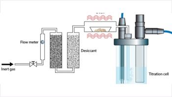
For successful use of the SEPPRO media, Genway provides a kit that has three buffers used for dilution–washing, stripping, and neutralization. The buffers are specified in the data sheet. Using the neutralization buffer, the media can be regenerated and used multiple times. An automated method for highly abundant protein partitioning of human serum and plasma samples before proteomic analysis was the subject of a recent paper (13). Using the experimental setup shown in Figure 7, the author was able to use a 52 mm × 7 mm SEPPRO MIXED12 flow-through column for 115 sample runs of 25 μL each of human serum/plasma.
Figure 7
Recently, Genway has developed another IgY microbead column called the SuperMix column. This column is designed to deplete the next level of serum and plasma proteins, the moderately abundant proteins (MAP). The SuperMix column was developed by immunizing chickens with a flow-through fraction of IgY-12 column and constructing the column with affinity-purified IgY antibodies against the flow-through proteins of IgY-12. This second column used in series with the IgY-12 column is said to allow one to look at additional low-abundance proteins that were masked in earlier studies.
The ProteoPrep 20
The ProteoPrep 20 Plasma Immunodepletion Kit, a product from Sigma Aldrich (St. Louis, Missouri), was introduced in 2005. The kit contains multivalent antibody affinity media in a spin column format and is designed to remove 20 proteins from human plasma. The ProteoPrep 20 media are based upon small recombinant single-chain antibodies and conventional antibodies specific for human plasma proteins that are bonded to agarose particles at a fairly high density. A poster presented at a National Cancer Institute Proteomic Technologies Workshop showed the depletion of 97–98% of total protein from plasma (14). The kit comes complete with the prepacked column (0.3 mL in a spin column format only), proprietary solutions including an equilibration buffer and an elution solvent and preservative concentrate for column storage. The column will accommodate an 8-μL injection of plasma and is said to allow at least 100 injections. During the first pass (initial depletion) about 90–95% of high-abundance proteins are removed. A plasma sample is incubated for 15–20 min with column media, and, after a centrifugation and washing step, the flow-through (depleted plasma) is collected. The combined flowthroughs from 10 runs are pooled, concentrated and redepleted (second pass through the resin) for a final depletion of >99% of targeted proteins. With a high removal of protein, more low-abundance proteins are observed because higher sample loads can be placed onto 2-D gels or HPLC columns (14).
Proteospin Abundant Serum Protein Depletion Kit
Unlike the other poducts in this category, the Proteospin Abundant Serum Protein Depletion Kit, supplied by Norgen Biotek Corporation (St. Catherines, Ontario, Canada) is not based upon affinity depletion but on an ion-exchange mechanism. Relative to the antibody-based products, one advantage of the ion-exchange approach to protein depletion is the cost of the product. Each spin column can deplete up to 500 μg of total serum protein. In their studies, Norgen found that albumin is depleted by 70%, alpha-antitrypsin by 90%, and transferrin and haptoglobin by 50%. Their column can handle serum and plasma samples from a variety of sources including human and various animals. With their experimental protocol, 10 samples can be processed in less than 30 min. In addition to the mini-spin columns, the kit consists of column activation and wash buffer, elution buffer, and a neutralizer solution along with some elution tubes.
Comparison of the Various Depletion Approaches
Although it is not the intent of this article to compare details and pros and cons of the various high-abundance protein depletion strategies, we will cover some of the relative tradeoffs in choosing which approach to pursue. The simplest approach protein precipitation is the least costly and most straightforward, but all proteins of high molecular weights are precipitated, leaving only the lower molecular weight ones (<20 kDa) in the supernatant. There is a high probability of coprecipitation of low-abundance proteins in the process. The time-tested dye affinity approach is also a low-cost approach and can reduce albumin to a lower level, but there is greater cross reactivity (lack of specificity) and potential loss of valuable medium-to-low-abundance proteins.
The single-antibody columns that are specific to IgG and HSA can capture a good percentage of their target protein but still leave behind many high-abundance proteins. The protein A, G, and L resins can remove much of the IgGs successfully but are more effective when used in combination with an HSA depletion affinity phase. Some of the more esoteric approaches that have been published such as peptide affinity and multilectin affinity can prove useful, but there are no commercial products packaged conveniently to take advantage of the technology. The Gradiflow and isoelectric trapping can be quite selective multidimensional techniques, but both require the purchase of a new instrument. Ion-exchange chromatography, when combined with size-exclusion chromatography or reversed-phase chromatography, does not provide enough selectivity, and low-abundance proteins can be coeluted with the high-abundance proteins and can be lost in the process.
The newest multiple affinity approaches are now getting the most attention. In general, they are more selective and with optimized solution chemistries minimize the sorption of nontargeted proteins. Overall, their sample capacities appear to be lower than some of the more classical approaches, but they remove anywhere from 4 to 20 high-abundance proteins. The flowthrough medium-to-low-abundance proteins can be collected by pooling multiple passes and concentration. Thus, very small concentrations of proteins never observed before can be uncovered in 2-D gels and by LC–MS. Previously, many of these proteins were masked by the high background of the high-abundance proteins and were lost in the process.
The HPLC column format lends itself to automation, particularly important when large numbers of samples are encountered. Fractions of both flowthrough proteins and eluted high-abundance proteins can be collected automatically and pooled using modern LC instrumentation capabilities. The more rigid base packings allow the media to withstand the higher pressures or flow rates that can be encountered in the use of the HPLC column format. Flowthrough UV detectors can follow the sorption and elution of the protein bands. Having the entire process automated leads to increased reproducibility and the ability to quantitate the experiments.
There has been no formal publication comparing all of the multiple-affinity and ion-exchange approaches covered in Table IV. The MARS configuration has been around the longest and, thus, has more published results (2,5,7–10,15–30,53), but the other products are starting to see some published applications so that results can be compared (4,31–33).
Conclusions
There are multiple approaches to the removal of one or more high-abundance proteins from human serum and plasma. The idea of removing these high-abundance proteins allows one to "deep mine" the low-abundance proteins that have escaped detection due to masking effects in 2-D and HPLC investigations. With the wide concentration range of medium-to-low abundance proteins that are still present in depleted serum and plasma samples, there are still many challenges ahead to identify and relate them to various disease states or for therapeutic investigations.
Peter Mrozinski joined H-P/Agilent in 1991 as a Mass Spectroscopy Application Scientist and is currently an Applications Scientist for BioSeparations and Reagents.
Nina Zolotarjova joined Agilent Technologies in 2002. As a senior R & D Scientist in Columns & Supplies Division, she concentrates her efforts on the development of new Proteomics Separation tools and technologies.
Ronald E. Majors "Sample Prep Perspectives" Editor Ronald E. Majors is business development manager, Consumables and Accessories Business Unit, Agilent Technologies, Wilmington, Delaware, and is a member of LCGC's editorial advisory board. Direct correspondence about this column to "Sample Prep Perspectives," LCGC, Woodbridge Corporate Plaza, 485 Route 1 South, Building F, First Floor, Iselin, NJ 08830, e-mail
References
(1) W.-C. Lee and K.H. Lee, Anal.Biochem. 324, 1–10 (2004).
(2) Bjorhall, T. Miliotis, and P. Davidsson, Proteomics 5, 307–317 (2005).
(3) J.M. Jacobs, J.N. Adkins, W.-J. Qian, Y. Shen, D.G. Camp III, and R.D. Smith, J. Proteom. Res. 4(4), 1073–1085 (2005).
(4) Y. Ogata, M.C. Charlesworth, and D.C. Muddiman, J. Proteom. Res. 4(3) 837–845 (2005).
(5) B.A. Chromy, A.D. Gonzales, J. Perkins, M.W. Choi, M.H. Corzett, B.C. Chang, C.H. Corzett, and S.L. McCutchen-Maloney, J. Proteom. Res. 3, 1120–1127 (2004).
(6) L. Govednik, P. Brne, B. Gabor , M. Barut, A. Strancar, and M. Strlic, Chromatographic Characterization of Affinity and Pseudoaffinity Columns for the Depletion of IgG and HSA from Plasma Samples, 12th International Symposium on Separation Sciences, Poster Paper P1, Lipica, Slovenia, Sept. 27–29, 2006.
(7) J. Martosella, N. Zolotarjova, H. Liu, G. Nicol, and B.E. Boyes, J. Proteom. Res. 4(5), 1522–1537 (2005).
(8) K. Zhang, N. Zolotarjova, G. Nicol, J. Martosella, L.-S. Yang, C. Czafranski, J. Bailey, and B.Boyes, Agilent Application Note, 2003.
(9) J. Brand, T. Haslberger, W. Zolg, G. Pestlin, and S. Palme, Proteomics 6, 3236–3242 (2006).
(10) Z. Shen, E.J. Want, W. Chen, W. Keating, W. Nussbaumer, R. Moore, T.M. Gentle, and G. Siuzdak, J. Proteom. Res. 5, 3154–3160 (2006).
(11) X. Fang, L. Huang, J.S. Feitelson, and W.-W. Zhang, Drug Discovery Today Technol. 1, 141–148 (2004).
(12) Seppro Protein Fractionation Services Brochure, Genway, San Diego, California.
(13) M. Lynch, Amer. Biotech. Lab. 37(5), 24–26 (2005).
(14) M. Schuchard, C. Melm, A. Crawford, H. Chapman, S. Cockrill, K. Ray, R. Mehigh, D. Chen, and G. Scott, "Specific Depletion of Twenty High Abundance Proteins from Human Plasma", NCI Proteomic Technologies Regents Resource Workshop, Chicago, Illinois, Dec. 11–12, 2005.
(15) G. Maccarrone, D. Milfay, I. Birg, M. Rosenhagen, F. Holsboer, R. Grimm, J. Bailey, N. Zolotarjova, and C.W. Turck, Electrophoresis 25, 2402–2412 (2004).
(16) S.Y. Cho, E.Y. Lee, J.S. Lee, H.Y. Kim, J.M. Park, M.S. Kwon, Y.K. Park, H.J. Lee, M.J. Kang, J.Y. Kim, J.S. Yoo, S.J. Park, J.W. Cho, H.S. Kim, and Y.K. Paik, Proteomics 5, 3386–3396 (2005).
(17) N. Zolotarjova, J. Martosella J, G.Nicol, J. Bailey, B.E. Boyes, and W.C. Barrett, Proteomics 5, 3304–3313 (2005).
(18) R.L. Moritz, A.B. Chippingdale, E.A. Kapp, J.S. Eddes, H. Ji, S. Gilbert, L.M. Connolly, and R.J. Simpson, Proteomics 5, 3402–3413 (2005).
(19) X. Li, Y. Gong, Y. Wang, S. Wu, Y. Cai, P. He, Z. Lu, W. Ying, Y. Zhang, L. Jiao, H. He, Z.Zhang, F. He, X. Zhao, and X. Qian, Proteomics 5, (2005).
(20) A.K. Yocum, K. Yu, T. Oe and I.A. Blair, J. Proteom. Res. 4(5), 1722–1731 (2005) .
(21) J. Wu, M. Kobayashi, E.A. Sousa, W. Liu, J. Cai, S.J. Goldman, A.J. Dorner, S.J. Projan, M.S. Kavuru, Y. Qiu, and M.J. Thomassen, Molec. Cellular Proteomics 4.9, 1251–1264 (2005).
(22) H. Wang, S.G. Clouthier, V. Galchev, D.E. Misek, U. Duffner, C.-K. Min, R. Zhao, J. Tra, G.S. Omenn, J.LM. Ferrara, and S.M. Hanash, Mol. Cell Proteomics 4, 618–625 (2005).
(23) H.Molina, J. Bukenborg, G.H. Reddy, B. Muthusamy, P.J. Scheel, and A. Pandey, Mol. Cell. Proteomics 4, 637–650 (2005).
(24) L.A. Echan, H.-Y. Tang, N. Ali-Khan, K. Lee, and D.W. Speicher, Proteomics 5, 3292–3303 (2005).
(25) H.-Y. Tang, Proteomics 5, 3329–3342 (2005).
(26) N. Zolotarjova, B. Boyes, J. Martosella, L.-S. Yang, G. Nicol, K. Zhang, C. Szafranski, and J. Bailey, In "Separation Methods in Proteomics," G.B. Smejkal and A. Lazarev, (Ed.) 2006.
(27) L. Anderson and C.L. Hunter, Mol. Cell. Proteomics 5.4, 573–588 (2006)
(28) D. Sitnikov, D. Chan, E. Thibaudeau, M. Pinard, and J.M. Hunter, J. Chromatogr. B 832, 41–46 (2006).
(29) D.E. Misek, R. Kuick, H. Wang, V. Galchev, B. Deng, R. Zhao, J. Tra, M. Pisano, R. Amunugama, D. Allen, A.K. Walker, J.R. Strahler, P. Andrews, G.S. Omenn, and S.M. Hanash, Proteomics 5, 3343–3352 (2005).
(30) P. He et al, Proteomics 5, 3442–3452 (2005).
(31) L. Huang, G. Harvie, J.S. Fietelson, K. Gramatikoff, D.A. Herold, D.L. Allen, R. Amunngama, R.A. Hagler, M.R. Pisano, W.W. Zhang, and X. Fang, Proteomics 5, 3314–3328 (2005).
(32) T. Liu, W.-J. Qian, H.M. Mottaz, M.A. Gritsenko, A.D. Norbeck., R.J. Moore, S.O. Purvine, D.G. Camp, and R.D. Smith. Mol. Cel. Proteom. EPUB (2006).
(33) M.D. Schuchard, C.D. Melm, A.S. Crawford, H.A. Chapman, S.L. Cockrill, K.B. Ray, R.J. Mehigh, W.K. Kappel, and G.B. Scot, Origins 21, 17–23 (2005).
(34) Y.Y. Wang, P. Cheng, and D.W. Chan, Proteomics 3, 243–248 (2003).
(35) N. Ahmed, G. Barker, K. Oliva, D. Garfin, K. Talmadge, H. Georgiou, M. Quinn, and G. Rice, Proteomics 3, 1980–1987 (2003)
(36) D.A. Colantonio, C. Dunkinson, D.E. Bovenkamp, and J.E. van Eyk, Proteomics 5 3831–3835 (2005).
(37) M. Ramstrom, C. Hagman, J.K. Mitchell, P.J. Derrick, P. Hakansson, and J. Bergquist, J. Proteom, Res. 4, 410–416 (2005).
(38) D. Hinerfeld, D. Innamorati, J. Pirro, and S.W. Tam, J. Biomolec. Techniques 15(3), 184–190 (2004).
(39) Affibody Application Note Depletion of Abundant HSA from Serum, Affibody, Bromma, Sweden.
(40) C. Greenough, R.E. Jenkins, N.R. Kitteringham, M. Pirmohamed, B.K. Park, and S. R. Pennington, Proteomics 4, 3107–3111 (2004).
(41) S. Fahnestock, Trends in Biochem. Sci. 5, 1567 (1987).
(42) M. Fountoulakis, J.-F. Juranville, L. Jiang, D. Avila, D. Roder, P. Jakob, P. Berndt, S. Evers, and H. Langen, Amino Acids 27, 249–259 (2004).
(43) Y. Shen, J. Kim, E.F. Strittmatter, J.M. Jacobs, D.G. Camp III, R. Fang, N. Tolie, R.J. Moore, and R.D. Smith, Proteomics, 5, 4034–4045 (2005).
(44) N.I. Govorukhina, A. Keizer-Gunnink, A.G.J. van der Zee, S. de Jong, H.W.A. de Brujn, and R. Bischoff, J. Chromatogr. A 1009, 171–178 (2003).
(45) B. Herbert and P.G. Righetti, Electrophoresis 21, 3639–3648 (2000).
(46) G.L. Mikios and R. Maleszka, Proteomics 1, 30–41 (2001).
(47) D.L. Rothemund, V.L. Locke, A. Liew, T.M. Thomas, V. Wasinger, and D.B.Rylatt, Proteomics 3, 279–287 (2003).
(48) H.-J. Lee, E.-Y. Lee, M.-S. Kwon, and Y.-K. Paik, Current Opinion in Chemical Biology 10, 1–8 (2005).
(49) A. K. Sato, D. J. Sexton, L. A. Morganelli, E.H. Cohen, Q.-L. Wu, G. P. Conley, Z. Streltsova, S.W. Lee, M.Devlin, D.B. DeOliveira, J. Enright, R.B. Kent, C.R. Wescott, T.C. Ransohoff, A.C. Ley, and R.C. Ladner, Biotechnol. Prog. 18, 182–192 (2002).
(50) T. Baussant, L. Bougueleret, A. Johnson, J. Rogers, L. Menin, M. Hall, P.-M. Aberg, and K. Rose, Proteomics 5, 973–977 (2005).
(51) T.C. Petric, P. Brne, B. Gabor, L. Govednik, M. Barut, A. Strancar, and L.Z. Kralj, J. Pharm. Biomed. Anal., (in press).
(52) W.-H. Jin, J. Dai, S.-J Li, Q.-C. Xia, H.-F. Zou, and R. Zeng, J. Proteom. Res. 4, 613–619 (2005).
(53) Z. Yang and W.S. Hancock, J. Chromatogr. A 1053 (1–2), 79–88 (2004).
(54) A.J. Alpert and A.K. Shukla, ABRF 2003, poster #P111-W.
(55) J. Zhang, D.R. Goodlet, E.R. Peskind, J.F. Quinn, Y. Zhou, Q. Wang, C. Pan, E. Yi, J. Eng, R.H. Aebersold, and T. J. Montine, J. Neurobiol. Aging 26, 207–227 (2005).
Articles in this issue
over 19 years ago
How Little Can I See?over 19 years ago
The Saga of the Electron-Capture Detectorover 19 years ago
The Future of Drug Lead Development and Analysisover 19 years ago
Peaks of Interestover 19 years ago
High Performance Liquid Chromatographyover 19 years ago
Application of SEC-MALS in Paint ManufacturingAdvertisement
Related Content
Advertisement

Webcasts


Advertisement
Advertisement
Trending on LCGC International
1
AI/ML In Practice: Predicting the Measurable Chemical Space for Nontargeted LC–ESI–HRMS Analysis Workflows
2
Centrifugal Partition Chromatography, and Why Purification Labs Are Paying Attention
3
Sensor GC Tests Water's Effect on Hangovers
4
Working Toward Cross-Lab Metabolomics Confidence
5