

News|Articles|September 7, 2025
Single to Triple: Fundamentals and Modes of Bench-Top Gas Chromatography-Triple Quadrupole Mass Spectrometry (GC–MS/MS)
Author(s)Nicholas H. Snow

Listen
0:00 / 0:00
Key Takeaways
- GC–MS/MS provides high resolution, selectivity, and sensitivity, enhancing traditional GC–MS with multidimensional detection capabilities.
- Triple quadrupole systems in GC–MS/MS enable selected reaction monitoring (SRM) and multiple reaction monitoring (MRM) for improved analysis.
Advertisement
Among chromatographic separation and detection methods, gas chromatography-tandem mass spectrometry (GC–MS/MS) offers an unparalleled combination of high resolution, selective, universal, and sensitive detection. MS/MS may easily be described as the ultimate detector for GC. In this installment, we will describe several modes of MS/MS detector operation. After a brief overview of terminology and fundamentals, we see that MS/MS can operate as a single quadrupole system, providing classical mass spectra and operating in both foll scan and selected ion monitoring modes. We will then explore several modes of multidimensional operation, including scanning and selecting ions in each of the two dimensions, focusing on selected reaction monitoring (SRM), which provides maximum selectivity and sensitivity.
In previous installments, we have discussed various aspects of traditional gas chromatography-mass spectrometry (GC–MS), including troubleshooting, instrumentation and the modes of data analysis: full-scan, extracted ion chromatograms, and selected ion monitoring (1–3). We have seen the versatility of GC–MS, especially that, as a single detector, it is both universal and selective. GC–MS provides both the blessing and the curse of detecting nearly all compounds that reach the ionization source. With selected ion monitoring, GC–MS easily provides quantitative analysis of liquid samples at ng/mL concentrations and lower, combined with qualitative analysis based on multiple ions or on the full mass spectrum.
Triple quadruple mass spectrometry was originally developed in the late 1970s and the first instruments were large and cumbersome; they could be easily interfaced to gas chromatography, but this was not common due to the complexity and expense. Bench-top triple quadrupole systems have been available since the 1990s, and, as the decades have progressed, have become simpler to operate and less expensive. While there is still a significant capital expense in addition to the cost of GC–MS, GC–tandem mass spectrometry (MS/MS) is now much more accessible to more laboratories.
In this article, we will discuss the modes of MS/MS as a detector for GC, and see the additional versatility and capabilities that multidimensional detection in MS/MS provides along with those for traditional single dimension MS. Although there are multiple systems and configurations for MS/MS, this article will focus on triple quadruple MS. A simple diagram of a triple quadrupole mass spectrometer is shown in Figure 1. The basic components are very similar to traditional single quadrupole MS. In GC–MS/MS, we use the same ionization sources as for GC–MS, usually classical electron impact or ionization. As in traditional GC–MS, ions formed in the ionization source ad passed to the mass analyzer through a series of lenses that focus the ions along the long axis of the quadrupoles. The first quadruple following the ionization source (usually labeled Q1) is operated by the same two modes as in traditional GC–MS: full scan, in which all masses are passed through, or selected ion monitoring, in which a number of user-selected ions are passed.
The second quadruple (Q2) provides re-focusing of the ions passed from Q1 and a venue for collision induced dissociation or re-ionization of the transmitted ions using a soft chemical ionization process. These newly generated ions are then transferred using lenses to Q3, which can be operated in either full scan or selected ion monitoring mode. Finally, the ions are detected by an electron multiplier, as in traditional GC–MS. Note that an MS/MS can be operated in single quadrupole mode by simply passing ions generated in the ionization source through Q1 and Q2 and operating Q3 as if it is a singe quadrupole.
Seeing two modes of operation for Q1 and two for Q3, we can consider several possibilities for mass analysis experiments using MS/MS. Figure 2 shows a simple diagram showing three possibilities, each of which offers benefits and difficulties, depending on the needed analysis.
The first possibility is to operate Q1 in selected ion monitoring mode, with one or a few ions chosen to pass into Q2 as precursor ions. In Q2, these precursor ions are then further dissociated by collision with a regent gas, termed collision induced dissociation. The product ions generated in Q2 are passed to Q3, which is operated in full scan mode. This mode is especially useful for qualitative analysis, spectral interpretation, and multiple reaction monitoring (MRM) method development.
For spectral interpretation we begin with a classical one-dimensional mass spectrum of the analyte, easily obtained using a Q3 scan. An ion of choice, the base peak or molecular ion is a good place to start, can then be selected for the Q1 selected ion monitoring. The resulting fragmentation in Q3 can then be used to assign smaller fragments in the original mass spectrum to the larger fragments from which they originate. This is a process of assigning the reactions and transitions that occur in the ionization source. This is an excellent way to confirm mass spectral interpretations performed according to classical rules such as those provided decades ago by McLafferty (4). This can be summarized as SIM → Scan and is termed a product scan.
In the second possibility, Q1 is operated full scan and all the ions with all the masses generated, are passed to Q2. These initially generated ions are termed precursor ions. Q2 is then used to select one (or a few) precursor ions for reionization and analysis by Q3. The ions passed to Q3 and ultimately detected by the electron multiplier are termed product ions. In the past, the terms parent ion and daughter ion were used to describe precursor and product ions; this newer terminology avoids gender. This technique is very similar to one dimensional selected ion monitoring and has similar performance characteristics. This is summarized as Scan → SIM and is termed a parent or precursor scan.
Finally, and perhaps most importantly for quantitative analysis, both Q1 and Q3 can be operated in SIM mode. This is termed multiple reaction monitoring (MRM), as several mass transitions or reactions can be observed simultaneously, as in SIM for single masses. Method development, using the first two modes is required, but once set up, we will see how MRM can provide extremely low detection limits and high selectivity. In the remainder of this installment, we will focus on MRM, with an example of how it works and why it provides such excellent sensitivity and selectivity. MRM can be summarized as SIM → SIM.
Let us now turn to a summary of how an MRM experiment is set up, which will also show why MRM is the gold standard for sensitivity in chemical analysis. Figure 3 shows a structure and mass spectrum of a common steroid, 17β-estradiol. Estradiol is perhaps the best-known steroid hormone, and it plays a role in many reproductive and metabolomic functions in the body. It is also known as an emerging contaminant in natural waters, as drugs containing estradiol are eventually excreted and passed into water systems (5,6).
17β-estradiol is a very well-known compound; if this were an unknown, the MRM transitions could be determined using a Q3 scan to find the classical mass spectrum and then product scans to determine the transitions. In this case, the transitions are known and are indicated on the figure.
Once the transitions are determined, usually three or four are used. One transition is used for quantitation and the others for confirmation, much in the same manner as in classical SIM, in which one is used for quantitation and the others for confirmation. If any ions in SIM, or transitions in MRM, are not detected, the analyte is not identified. If all ions or transitions are present, identification is considered positive.
Table I shows transitions and optimized MRM parameters for several steroids in a mixture. Full details about the experimental conditions can be found in Reference 5, which is available open access. To maximize sensitivity, the collision energy on Q2 for each analyte can be optimized separately. This is a simple experiment involving varying the collision energy to maximize the peak height or area. In this experiment, we note the variation in collision energy from a low of 10 eV to a high of 20 eV. We see that this energy is much lower than the energy in the election ionization source, which is generally set at 70 eV. This low collision energy limits fragmentation, making Q3 spectra (following Q1 analysis) simpler than classical one-dimensional spectra.
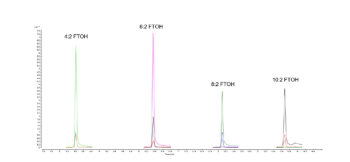
Finally, with the transitions and collision energies programmed into the system, an MRM experiment can be performed. The sample preparation and chromatography are the same as in classical GC–MS, in this experiment, solid phase microextraction (SPME) was used for sample preparation and injection and a classical 5% phenyl polydimethyl siloxane capillary column was used for the separation. Figure 4 shows the MRM chromatograms for the several steroids shown in Table I at 1 ng/mL (1 ppb) concentration in water. We see very high signal to noise ratios for all compounds except diethylstilbestrol, likely an artifact of the extraction. For this mixture, the detection limits using MRM were sub part-per-trillion for most of these steroids.
Why do we see such low detection limits and high sensitivity with MRM when the mass injected into the column and reaching the detector is the same in classical GC–MS? This is explained by considering both signal and noise; the signal to noise ratio. If we look back at Figure 3, the full scan spectrum, we see that there are many small peaks and if we able to look closely enough, we would see a positive signal for every mass, whether it appears in the analyte’s mass spectrum or not. This is classical detector noise.
In classical SIM, we only allow a single mass of choice to pass to the detector, eliminating the noise from all the other masses in the spectrum. While the signal is no larger, the noise is significantly reduced, accounting for the greater sensitivity. In MRM, we extend this idea an additional step, noise is further suppressed, resulting in much higher signal to noise ration than in SIM, and very low detection limits.
If you are already using GC–MS, you know it to be both universal and selective, sensitive and versatile. There are few problems in gas chromatography that cannot be solved by GC–MS, but two of them are interpreting complex unknown spectra and obtaining ultra-low detection limits, for compound such as emerging contaminants in the environment. Using a similar bench-top platform, GC–MS/MS can address both problems. Product scans can be used to assist in both spectral interpretation and in MRM method development. MRM can be used for ultra-trace quantitative analysis, with effective sample preparation and chromatography allowing detection limits of part-per-trillion or lower. Triple quadrupole mass spectrometry provides the gold standard detector for gas chromatography.
References
- Mizvesky, J.; Snow, N. H. Fundamentals of Benchtop GC–MS Data Analysis and Terminology. LCGC Intern.2025, 2 (3), 12-14, DOI: 10.56530/lcgc.int.sk6076k5
- N.H. Snow, N. H. Stopping GC and GC–MS Problems Before They Start. LCGC North Am. 2019, 37 (1), 18-23.
- Snow, N. H. Flying High with Sensitivity and Selectivity: GC–MS to GC–MS/MS. LCGC North Am. 2021, 39 (2), 61-67. DOI: 10.56530/lcgc.na.yn3065q6
- McLafferty F. W.; Turecek, F. Interpretation of Mass Spectra, 4th Edition; University Science Books, 1993.
- Chopra, S.; Gomes, P. F. C. L.; Dhandapani, R.; Snow, N. H. Scientia Chromatographica. 6(2), 105–116. (2014). https://www.iicweb.org/scientiachromatographica.com/files/v6n2a02.pdf
- Chopra, S. Extending the Limits of Solid-phase Microextraction, PhD Dissertation, Seton Hall University, 2014.
About the Author
Advertisement
Related Content
Advertisement


Advertisement
Advertisement
Trending on LCGC International
1
The Silent Crisis: Can The Demise of Chromatography Teaching in Universities Be Reversed?
2
Multiplexed HILIC-HRMS Method Quantifies LPC and CAR Biomarkers for HCC
3
SPME in Practice: Techniques, Tips, and Troubleshooting
4
HTC-19 Insights: HPLC–XRF for Environmental Analysis — Future Developments
5