
|Articles|April 6, 2021
- April 2021
- Volume 17
- Issue 04
Tips & Tricks GPC/SEC: Separation Range and Resolution
Author(s)Daniela Held, Wolfgang Radke
Advertisement
Whereas the elution time in interaction chromatography can be widely varied by applying a suitable eluent composition or gradient slope, the separation in GPC/SEC is restricted by the interstitial and the void volumes of the column. At the same time, the molar mass separation range of a GPC/SEC column or column combination is restricted by the column’s pore size distribution. Because GPC/SEC is an isocratic method GPC/SEC resolution cannot be tuned by gradient slope, nor does eluent selection provide many options to optimize separation. Therefore, a proper choice of the GPC/SEC column or column combination is mandatory to achieve high resolution separations in GPC/SEC. This instalment describes the interplay between column length, pore size distribution, and particle size to optimize GPC/SEC separations.
In macromolecular science, GPC/SEC is a major separation technique. GPC/SEC separates individual macromolecules based on their hydrodynamic sizes in solution. A mixture of macromolecules of different sizes is injected into the chromatographic column, filled with porous particles. As the macromolecules migrate through the column, they pass pores of different sizes. Molecules larger than the pores cannot diffuse into the pores, while for molecules much smaller than the pores there is no substantial differentiation. With a size comparable to the pore size, macromolecules can migrate into the pores and are separated based on their residence time in the pores, which depends on their size in solution. Since macromolecules of the same chemical structure but varying in molar masses exhibit different sizes, they can be separated by GPC/SEC according to molar mass.
It then follows that a column providing only one pore size can only separate a limited molar mass range. As a rule of thumb, a single pore size column allows separating macromolecules differing in molar mass by approximately two orders of magnitude (1,2,3). If samples of broad molar mass distribution have to be separated, either columns providing a mixture of pore sizes or combinations of columns having different pore sizes need to be applied.
While the molar mass range that can be separated by GPC/SEC is governed by the pore size distribution of the column packing, the resolution of two molar masses to be separated is additionally influenced by the total pore volume of the column or column combination and the particle size. The latter parameter needs to be taken into account, as it affects band broadening.
The first part of this instalment of Tips & Tricks will discuss the effect of pore size, pore size distribution, and column length on the ideal separation of two molar masses, while the second part will elucidate the effect of band broadening, in particular due to particle size on the resolution in GPC/SEC.
Effect of Pore Size on Molar Mass Separation Range
As mentioned above, the molar mass range that can typically be separated on a single pore size column covers approximately two orders of molar mass. Since the molar mass distribution of typical macromolecular samples often extends over several orders of magnitude (4), the column’s upper exclusion limit or the low molar mass separation limit might be reached if the molar mass range of the single pore size column is not well adjusted to the molar mass range of the sample.
Figure 1 schematically depicts the effects of selecting different single pore size columns to the separation of a broadly distributed sample. On the column characterized by the black calibration curve, the sample is well separated, as the whole molar mass distribution is covered by the nearly linear range of the calibration curve. In contrast, on the column characterized by the blue calibration curve, corresponding to a column of smaller pore diameter, the sample reveals a rather narrow high molar mass peak followed by well separated oligomers. The steep increase at low elution volume is due to the very high molar mass fraction of the material approaching the exclusion limit of the column. Because the high molar mass fraction is not effectively separated any longer, the high molar mass molecules elute in a very narrow elution volume range, piling up their concentrations which finally results in a sharp exclusion peak. Such exclusion effects can result in additional shoulders or even additional peak maxima, which are, however, not a characteristics of the sample’s molar mass distribution, but originate from an unsuited molar mass separation range of the column. The chromatogram obtained on the column characterized by the red calibration curve reveals a well-separated high molar mass fraction, while the oligomers are not resolved and elute as a non-separated single peak. The lack of separation of the oligomers results from the steepness of the calibration curve at high elution volumes, which render the oligomers to elute very close to each other thereby merging into a single peak. It should be noted that the molar mass corresponding to the weight average molar mass of the sample is within the separating range of each of the three calibration curves. Thus, selecting a column based on published calibration curves considering only the weight average molar mass of the sample, without taking into account the width of the molar mass distribution, can result in selecting an unsuitable column.
Effect of Pore Size Distribution
The width of the molar mass ranges covered by typical macromolecular samples in conjunction with limited separation range of a single pore size, often necessitates the provision of not only one specific pore size but a distribution of pore sizes. Providing a wider range of pore sizes in a column or column combination extends the molar mass range that can be separated. Columns containing not only a single pore size, but a range of different pore sizes are referred to as “mixed bed”, “linear”, or “multipore” columns.
Since a single column packed with particles with a broad pore size distribution extends the molar mass range that can be separated, but does not alter the elution volume range over which the separation occurs, a steeper calibration curve results, as compared to a single pore size column. This is evident in Figure 2.
Here the two molar masses indicated will be separated by a lower elution volume range on a mixed bed column of broad pore size distribution, as compared to the separation on a column providing only a single pore size.
Effect of Column Length
Instead of packing a single column with a material of broad pore size distribution, it is possible to couple several columns, each of which having a narrow pore size distribution. Such a column combination or column bank provides a large molar mass separation range, due to the different pore sizes present. In addition, such a column bank has a large pore volume. Thus, the volume range over which the separation takes place is extended as compared to a single column filled with the same pore size mixture (mixed bed column). In both approaches the same pore size distribution and thus the same molar mass separation range exists. However, the separation volume for the column bank is larger than of the single column. Therefore, the slope of the calibration curve is less steep for the former, and two molar masses elute further apart, as on a single column. This is visualized in Figure 3. This enhanced separation is achieved, however, at the cost of longer analysis times and larger solvent consumption.
It is also possible to couple several linear or mixed bed columns to a column bank, maintaining the same separation range and increasing the separation volume, thereby decreasing the slope of the calibration curve. However, mixed bed or linear columns are usually not well adapted to the molar mass distributions of the specific samples to be analyzed but are designed as “general purpose” columns. Therefore, linear or mixed bed columns often do not only provide pores required for the specific separation problem but also pore sizes which are of no particular advantage, due to being either too large or too small. Since these pores contribute to the total pore volume, linear or mixed bed columns usually increase analysis time and solvent consumption without providing the full benefit regarding sample separation.
Effect of Particle Size
As mentioned above, chromatographic resolution is determined by two factors: the difference in elution volume between the two components that need to be separated and by band broadening effects, which deteriorate the separation of the two ideally separated peaks.
While the spacing between the different components in GPC/SEC is determined by
the slope of the calibration curve and the available column volume, as explained above, several factors contribute to band broadening, for example, a finite injection and detector volume, broadening/mixing within the capillaries and detectors, and column band broadening. In general, smaller particles reduce column band broadening. Band broadening effects are typically quantified by injecting a monodisperse compound and determining the plate count (Nth) from peak width (w1/2) and elution volume at peak maximum (Vp), with L being the column length in cm:
Due to the different diffusion and transport phenomena occurring in a chromatographic system, band broadening and thus plate counts depend on variety of parameters. Despite this, it is good practice to determine the number of theoretical plates regularly under identical conditions as it provides an easy tool to track the system performance. A significant decrease in plate count might indicate problems with the instrument and/or the separating column. However, as mentioned before, plate count is but one factor to achieve good GPC/SEC resolution. The other factor is the slope of the calibration curve (5).
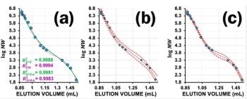
The interplay of these parameters is schematically shown in Figure 4. Here we investigate the separation of two molar masses present in the sample. When comparing the elugrams resulting from a column providing a flat (a) and a steep calibration curve (b) at the same instrument performance (peak width) the resolution between the two peaks is clearly higher for the column of lower slope. This is due to the distance between the two peaks being higher on the column providing the lower slope.
If we compare the chromatograms resulting from columns of identical slope but different plate counts (b, c), the resolution between the two peaks is diminished for the column of lower plate count (broader peaks, c). Such broader peaks might either result from aging of the column, which might lead to increased peak broadening, or from two columns having the same pore size distribution but differing in particle size.
It is not uncommon for SEC/GPC users to conclude from a low slope of the calibration curve to the existence of a well separating GPC/ SEC system. However, the above discussion showed that the slope of calibration curve is but one factor determining resolution. Some national and international standards stipulate a minimum for the specific resolution within the required separation range, which can only be realized using column combinations. The calculation of the specific resolution takes into account both, the slope of the calibration curve and the influence of band broadening. More details can be found in the Tips & Tricks instalment on “How to Test GPC/SEC columns” (5).
As mentioned above, lower particle sizes are beneficial from the perspective of band broadening. However, macromolecules, especially of high molar mass are prone to shear. Shear might result in stretching of the molecules and lead to elution, not determined any longer by the coil size of the macromolecule. Therefore, high molar mass polymers should be analyzed by GPC/SEC using larger particles. On the other hand, if oligomers or samples producing narrow peaks such as proteins need to be separated, band broadening should be reduced as far as possible, to obtain good resolution. Thus, lower particle size columns are recommended. The effect of particle size on band broadening and thus on oligomer separation is shown in Figure 5. Clearly the oligomer resolution is better the smaller the particles are.
Large particles are preferred for the separation of high molar mass molecules, whereas smaller particles are preferred for the resolution of oligomers. This seems to suggest the columns should be filled with mixtures containing both, large particles with large pores and also small particles with small pores, if broad molar mass samples need to be analyzed.
However, instead of combining the advantages by packing small particles with small pores and large particles with large pores in a single column or column combination, such an approach combines the drawbacks of both, as discussed in a previous instalment of Tips & Tricks for GPC/SEC (6).
Summary
- Resolution in GPC/SEC depends on the slope of the calibration curve and on plate count.
- The distance between two molar masses separated by GPC/SEC depends on the slope of the calibration curve.
- The molar mass range that can be separated by a single pore size column in GPC/SEC is restricted.
- Broad pore size distributions of a column’s packing material provide a large molar mass separation range at the cost of a steeper calibration curve, as compared to an individual pore size column.
- The identical molar mass separation range obtained using a column bank provides a less steep calibration curve, thus, larger elution volume differences.
- Separations for large macromolecules require large pore and particle sizes.
- Oligomer separations should be performed on columns providing small pore and particle sizes.
- Column combinations of large and small particles are not recommended for separating samples containing small and large molecules.
References
1. E.F. Casassa, J. Polym. Sci. B. Polym. Letters, 5(9), 773–778 (1967).
2. E.F. Casassa and Y. Tagami, Macromolecules 2(1), 14–26 (1969).
3. W. Radke, Macromol. Theory Simul. 10(7), 668–675 (2001).
4. W. Radke and D. Held, The Column 12(22), 19–22 (2016).
5. F. Gores and D. Held, The Column 10(14), 7–10 (2014).
6. D. Held and W. Radke, The Column 16(8), 17–21 (2020).
Daniela Held studied polymer chemistry in Mainz, Germany. She works in the PSS software and instrument department, and is also responsible for education and customer training.
Wolfgang Radke studied polymer chemistry in Mainz, Germany, and Amherst, Massachusetts, USA, and is head of the PSS application development department. He is also responsible for instrument evaluation and for customized training.
E-mail:
Website: www.pss-polymer.com
Articles in this issue
over 5 years ago
Vol 17 No 4 The Column April 2021 Europe & Asia PDFover 5 years ago
Vol 17 No 4 The Column April 2021 North American PDFover 5 years ago
Quantifying AAV Quality Attributes Using SEC–MALSover 5 years ago
Rising Stars of Separation Science: Mariosimone Zoccaliover 5 years ago
Analytical Laboratory Training: Far From Ideal!over 5 years ago
The LCGC Blog: Celebrating HPLC Pioneer Elmar Pielover 5 years ago
LCGC Europe and HTC-17 Launch 2022 HTC Innovation AwardAdvertisement
Related Content
Advertisement


Advertisement
Advertisement