
|Articles|July 1, 2016
- The Application Notebook-07-01-2016
- Issue 4
2D-LC–MS Characterization of Charge Variants Using Ion Exchange and Reversed-Phase Chromatography
Author(s)Sonja Schneider
Advertisement
Dr. Sonja Schneider, Agilent Technologies Inc.
This application note demonstrates the analysis of charge variants of Rituximab (both innovator and biosimilar molecules) using multiple heart-cutting two-dimensional liquid chromatography (2D-LC) with mass spectrometry (MS) detection.
Introduction
Monoclonal antibodies (mAbs) represent a class of highly advanced, but expensive, pharmaceutical products. The increasing demand for these biopharmaceuticals has led to the development of less costly biosimilars (1). To qualify the charge variant pattern of the mAbs, identification with mass spectrometry (MS) is necessary. However, identification using online MS analysis after ion exchange chromatography is not a straightforward procedure. This is mostly because of the incompatibility of the most common ion exchange buffers with the electrospray ionization process.
The Agilent 1290 Infinity 2D-LC solution enables automated desalting, and offers denaturation and, if necessary, extra separation by the addition of reversed-phase chromatography in the second dimension.
Experimental Conditions
1D - Mobile phase: A: 10 mM ammonium formate pH 6.2
1D - Mobile phase: B: 500 mM ammonium formate, pH 6.2
2D - Mobile phase: A: water + 5% FA
2D - Mobile phase: B: acetonitrile with 5% FA
1D flow rate: 0.2 mL/min
1D gradient: 0 min - 0% B
30 min - 40% B
35 min - 100% B
38 min - 0% B
1D stop time: 75 min
2D parameter mode: Heart-cutting
2D gradient stoptime: 3.0 min
2D cycle time: 4.5 min
Flow: 1 mL/min
Idle flow: 0.10 mL/min
2D gradient: 0.00 min - 10% B
2.5 min - 60% B
2.75 min - 90% B
Injection volume: 6 μL
Thermostat autosampler: 10 °C
Column temp.
1D column: 22 °C
Column temp.
2D column: 70 °C
DADs: 280 nm, 4 nm, Ref. 360 nm, 100 nm
Peak width 1D: 0.025 min (0.5 s response time) (10 Hz)
Peak width 2D: > 0.0063 min (0.13 s response time) (40 Hz)
Results
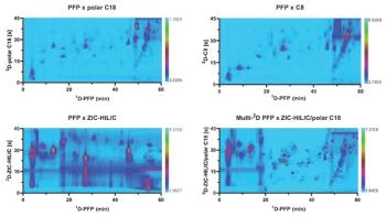
Figure 1 shows the results of the multiple heart-cutting 2D-LC analysis presented in the 2D-LC heart-cut viewer.
Figure 2 shows the deconvoluted spectra of charge variant main peaks 1–3 (Figures 2a–c). The mass of the first biosimilar main peak for G0F/G0F is 147,082 Da, identical to G0F/G0F of the innovator. The G0F/G0F of the second (Figure 2b) and third (Figure 2c) charge variant peak are shifted with an interval of about 128 Da, representing the mass of lysine. This leads to the assumption that the three main peaks are C-terminal lysine variants.
Conclusion
Innovator and biosimilar Rituximab charge variants were analyzed using weak cation exchange chromatography. Direct qualification of the charge pattern using online MS detection was enabled using a two-dimensional workflow with reversed phase in the second dimension for straightforward MS detection.
For research use only, not for use in diagnostic procedures.
Reference
- X. Hongwei, mAbs2(4), 379–394 (2010).
Agilent Technologies, Inc.
5301 Stevens Creek Blvd., Santa Clara, California 95051, USA
Tel: (800) 227 9770
Website:
Articles in this issue
about 10 years ago
Propylene Glycol (PG) in Alcoholic Beveragesabout 10 years ago
Analysis of Artificial Sweeteners by HILIC–MS Methodabout 10 years ago
Antibody Drug Conjugate (ADC) Analysis with SEC–MALSAdvertisement
Related Content
Advertisement


Advertisement
Advertisement