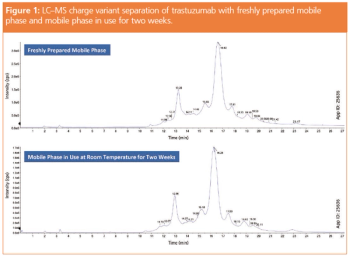
Biopharmaceuticals and Protein Analysis

Latest News


LCGC3_i1.jpg

Glycan isomer expressions have not been well studied, due to inefficient separation and structural identification techniques. Fortunately, with the development of novel separation techniques and liquid chromatography–mass spectrometry (LC–MS) based glycan isomer identification strategies, new efforts have been made to investigate the glycan isomers in various diseases. Here, we review the recent advances of several isomeric separation techniques for both N- and O-linked glycans.

figrue 6 L.png

Monoclonal antibodies (mAbs) are being developed at an explosive rate and have attracted great interest from both smaller biotech firms and big pharmaceutical companies. Developing mAbs and next-generation antibody–drug conjugates (ADCs) is highly demanding in many ways. From an analytical perspective, handling mAbs and ADCs presents many new challenges. This article describes how size-exclusion chromatography (SEC) combined with high-resolution mass spectrometry (HRMS) can be applied to the detailed characterization of mAbs and ADCs.

figure 1 L.png

Characterization of mAbs and related products requires the identification of chromatographic peaks with MS. However, the conventional salt- and pH-gradient elution techniques used in IEX are inherently incompatible with MS. Ammonium acetate- and ammonium carbonate-based mobile phase systems have been recently applied in IEX-MS, but the influence of the eluent composition on peak shape and retention has not been discussed nor studied systematically until now. The aim of the present study was to understand the impact of ionic strength, buffer capacity, and pH-response on the retention behaviour and peak shape of mAb species.

figure 1 L1552484426198.png

CZE–ESI‑TOF‑MS for the characterization of the mAb infliximab and its variants is presented. Infliximab was analyzed using a middle-up approach involving either reduction or digestion with the enzyme IdeS. A multilayer capillary coating of PB-DS‑PB in combination with a background electrolyte of 40% acetic acid provided efficient separation of the obtained antibody fragments, that is, LC and HC, as well as F(ab’)2 and Fc/2 parts. C-terminal lysine variants were also resolved. Recorded mass spectra of HC and Fc/2 fragments permitted assignment of 13 glycoforms and provided a quantitative profile, with G0F the most abundant glycoform (~50%). CZE–ESI-TOF-MS represents an efficient means for the straightforward analysis of a monoclonal antibody and its proteoforms.

figure 1 L.png

This article presents two case studies regarding the characterization of protein-DNA complexes using two complementary multi-angle light scattering (MALS) techniques, namely size-exclusion chromatography (SEC–MALS) to determine absolute molar mass of each component, and composition-gradient MALS (CG–MALS) to quantify stoichiometry and affinity at binding sites in solution.

figure 1 L1542213592344.png

Within the broad scope of analytical techniques required to characterize a protein, chromatographic methods have shifted towards high-flow analyses that can drop development time significantly. However, fast analytical methods for charge heterogeneity have lagged in development because current column technologies are ultrahigh-pressure liquid chromatography (UHPLC)-incompatible. This article will demonstrate the development of a high-flow method for charge variant analysis made possible through a bioinert titanium column flow path.

LCGCCTMS1_i1.gif

Among all the analytical techniques available for epitope mapping studies, hydrogen–deuterium exchange mass spectrometry (HDX-MS) is usually the fastest and easiest to carry out. We present here the epitope mapping of three distinct monoclonal antibody (mAb) candidates targeting the same antigen, an interleukin receptor. The goal is to establish the binding mode of these mAbs, and explain possible differences observed for in vitro binding and in vivo function.

figure 1 L.png

Glycosylation is a critical quality attribute (CQA) that can impact on product safety and efficacy of protein biopharmaceuticals. Characterization of N-glycans is therefore of paramount importance for the pharmaceutical industry. Hydrophilic interaction liquid chromatography (HILIC) combined with fluorescence detection (FLD) and 2-aminobenzamide (2-AB) labelling is the golden standard for the analysis of N-glycans enzymatically liberated from biopharmaceuticals. However, for phosphorylated N-glycans, that is, those attached on lysosomal enzymes, irreproducible data and recovery issues are observed on conventional liquid chromatography (LC) instrumentation and columns, which can be attributed to the interaction of the phosphate moieties with stainless steel components in the flow path. This article demonstrates the analysis of phosphorylated glycans with full recovery on a bio-inert LC system and PEEK-lined HILIC column.

Much of the conventional wisdom regarding size-phase separations of proteins has been negated thanks to development of superior chemistries and advances in research. In this article, details that the authors have found to be especially beneficial in achieving effective SEC separations are examined.